Case 42

 Clinical Presentation
A 39-year-old woman with multiple urinary tract infections.
 Imaging Findings

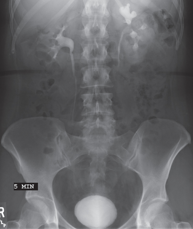
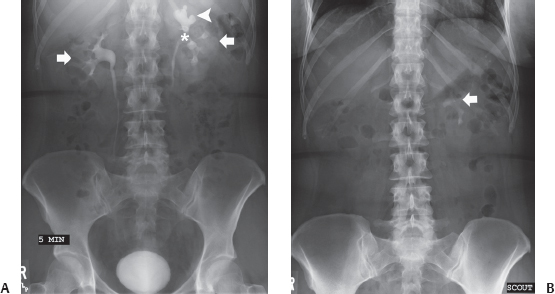
(A) A single 5-minute intravenous urogram (IVU) image shows that both kidneys (arrows) are normal in size, shape, position, orientation, outline, and parenchymal thickness. Mild caliceal blunting and dilatation (arrowhead) are seen on the left. The left renal pelvis (asterisk) is of normal size. The right collecting system and visualized portions of both ureters are normal. The urinary bladder is not fully distended but appears normal. (B) The scout image from the same IVU shows a staghorn calculus (arrow) in the left collecting system that was obscured by excreted contrast in the subsequent intravenous pyelogram (IVP) images.
 Differential Diagnosis
• Mild left hydronephrosis due to a stone:
Stay updated, free articles. Join our Telegram channel

Full access? Get Clinical Tree


