Case 43
Case History
A 72-year-old woman status post-right lumpectomy and radiation therapy has developed a new mammographic finding near the surgical site.
Physical Examination
• right breast: subareolar scar
• left breast: normal exam
Mammogram
Mass (Fig. 43–1)
• margin: spiculated
• shape: irregular
• density: equal density
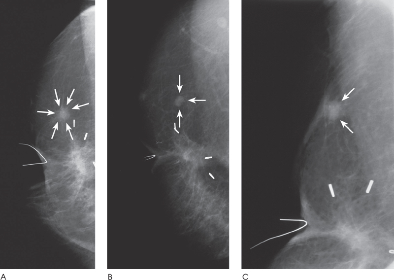
Figure 43–1. There is a spiculated density deep to the right nipple. This density corresponds to the scar from the patient’s lumpectomy. In the upper outer breast, there is a spiculated mass (arrows), which is separate from the main scar. This spiculated mass is new since the patient’s previous exam. (A). Right LM mammogram. (B). Right CC mammogram. (C). Right mammogram tangential to spiculated mass (arrows).
Ultrasound
Frequency
• 10 MHz
Mass


Stay updated, free articles. Join our Telegram channel

Full access? Get Clinical Tree


