Case 44
Case History
A 49-year-old woman presents with a new density on her right screening mammogram.
Physical Examination
• normal exam
Mammogram
Mass (Fig. 44–1)
• margin: indistinct
• shape: irregular
• density: equal
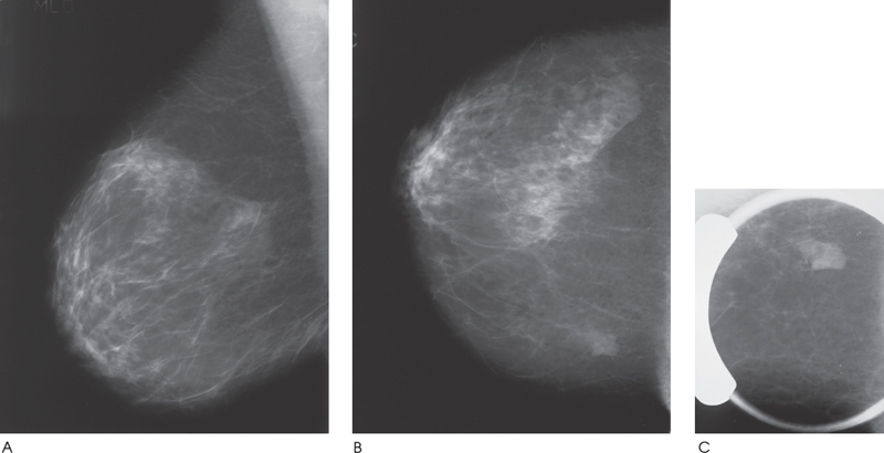
Figure 44–1. In the right upper medial breast there is an irregular mass that is partially obscured by the dense overlapping breast tissue. (A). Right MLO mammogram. (B). Right CC mammogram. (C). Right CC spot compression mammogram.
Ultrasound
Frequency
• 7.5 MHz
Mass
• margin: ill defined
• echogenicity: hyperechoic
• retrotumoral acoustic appearance: posterior shadowing distal to mass


Stay updated, free articles. Join our Telegram channel

Full access? Get Clinical Tree


