Case 44
 Clinical Presentation
A 38-year-old woman with chronic recurrent right flank pain. Computed tomography was performed.
 Imaging Findings
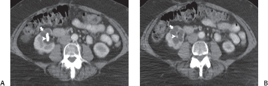
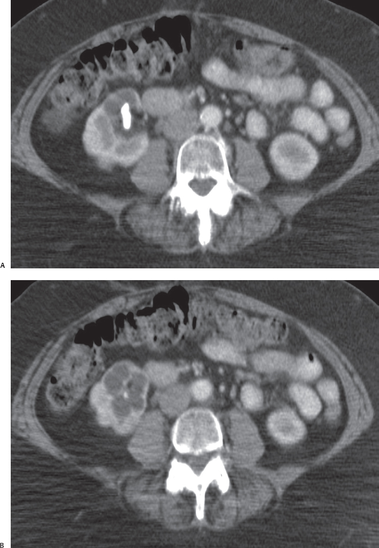
(A) Contrast-enhanced computed tomography (CT) image at the level of the kidneys shows a staghorn-type stone (arrowhead) in the right renal sinus. Some low-attenuation tissue (arrow) surrounds the stone. (B) Contrast-enhanced CT image at a level below that of Figure A shows the lower portion of the right renal staghorn stone (arrowhead). The area of low attenuation (arrow) is shown to be a mass of low attenuation value consisting of caliceal dilatation and replacement of the renal parenchyma at the lower pole of the right kidney.
 Differential Diagnosis
• Focal xanthogranulomatous pyelonephritis (XGP):
Stay updated, free articles. Join our Telegram channel

Full access? Get Clinical Tree


