Case 45
 Clinical Presentation
A 67-year-old man with a known history of prostate cancer develops some left lower limb swelling.
 Imaging Findings
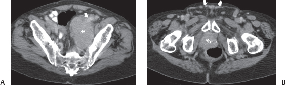
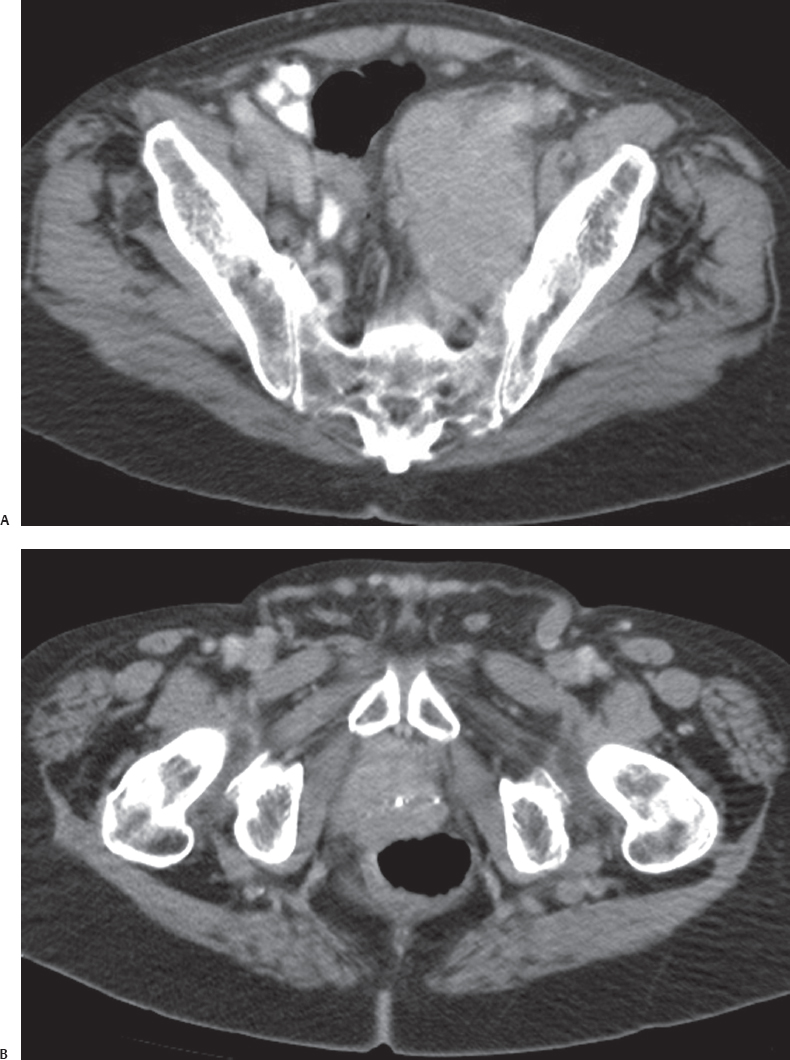
(A) Contrast-enhanced computed tomography (CT) image of the pelvis shows a large lobulated mass (asterisk) in the pelvis on the left side. The left external iliac vein (arrow) is partially encased by the mass. (B) Contrast-enhanced CT image at the samelevel shows a fairly normally sized prostate (asterisk) with some calcifications. There are multiple venous collaterals (arrows) in the subcutaneous tissue in front of the symphysis, suggesting obstruction of the left external iliac vein.
 Differential Diagnosis
Stay updated, free articles. Join our Telegram channel

Full access? Get Clinical Tree


