Case 48
Case History
A 63-year-old woman presents for a screening mammogram.
Physical Examination
• normal exam
Mammogram
Mass (Fig. 48–1)
• margin: spiculated
• shape: irregular
• density: equal density
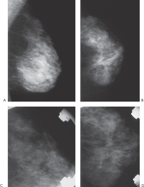
Figure 48–1. In the left upper medial breast, there is a spiculated mass that is more prominent on the CC view compared with the MLO or ML views. (A). Left MLO mammogram. (B). Left CC mammogram. (C). Left ML spot compression mammogram. (D). Left, CC spot compression mammogram.
Ultrasound
Frequency
• 11.5 MHz
Mass (Fig. 48–2)
• margin: ill defined
• echogenicity: hypoechoic
• retrotumoral acoustic appearance: severe shadowing, mass completely obscured
• shape: irregular (Fig. 48–2)


Stay updated, free articles. Join our Telegram channel

Full access? Get Clinical Tree


