Case 48
 Clinical Presentation
A 63-year-old man with difficulty voiding.
 Imaging Findings
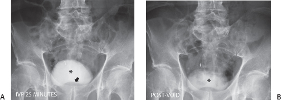
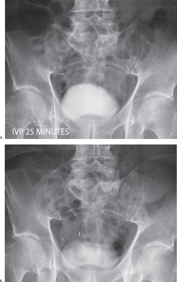
(A) A 25-minute intravenous pyelogram (IVP) image of the pelvis shows the bladder (asterisk) to be full. The outline is smooth, and there is a smooth, dome-shaped impression at the bladder base (arrow). (B) Postvoid IVP image of the pelvis. There is a significant amount of residual urine in the bladder (asterisk).
 Differential Diagnosis
• Enlarged prostate due to benign prostatic hyperplasia: An enlarged prostate with a smooth outline and obstructed emptying of the urine are suggestive of this entity.
Stay updated, free articles. Join our Telegram channel

Full access? Get Clinical Tree


