CASE 5 A 25-year-old man presents with fever and right upper quadrant pain. Fig. 5.1 (A) Utrasound of the abdomen shows a heterogeneous hypoechoic lesion in the right lobe of the liver (arrow). (B) Contrast-enhanced CT in the same patient shows a hypodense lesion with multiple enhancing septa ( arrow). Ultrasound of the abdomen (Fig. 5.1A) demonstrates a heterogeneous hypoechoic lesion in the right lobe of the liver with posterior acoustic enhancement. The lesion is hypodense on contrast-enhanced computed tomography (CT) (Fig. 5.1B) with multiple enhancing septa. No other lesions are seen. Pyogenic liver abscess Pyogenic hepatic abscesses have become less common due to early recognition and treatment of sepsis related to abdominal inflammatory processes, such as appendicitis and diverticulitis. In fact, no obvious source is identified in many patients with liver abscesses. Most infections are polymicrobial, with Escherichia coli
Clinical Presentation
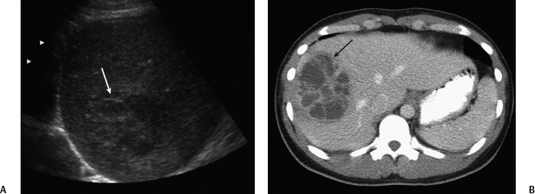
Radiologic Findings
Diagnosis
Differential Diagnosis
Discussion
Background
![]()
Stay updated, free articles. Join our Telegram channel


Full access? Get Clinical Tree


