Case 50

 Clinical Presentation
A 62-year-old man with immunocompromise and backache.
 Imaging Findings

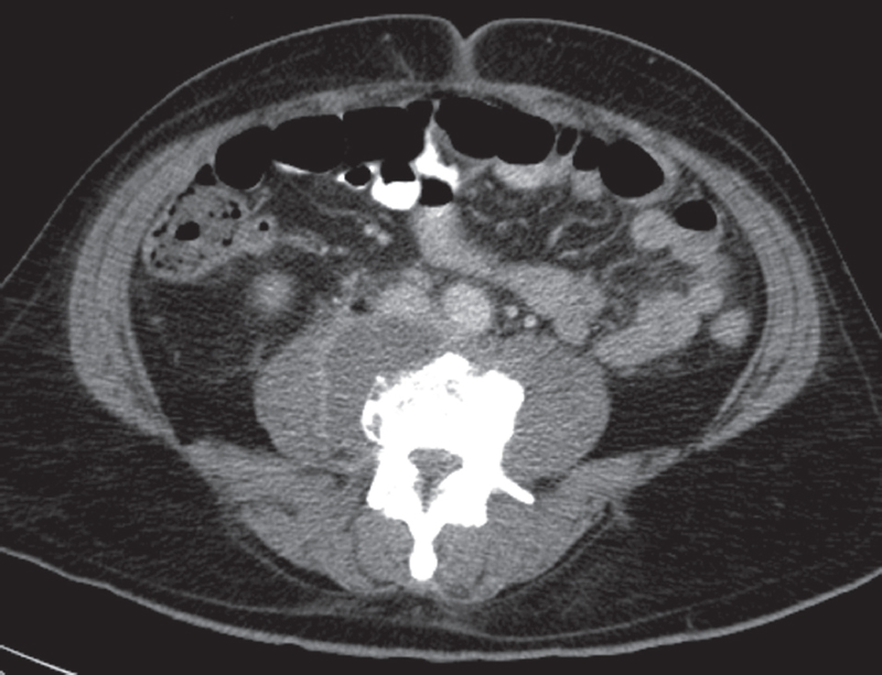
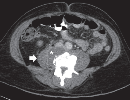
Contrast-enhanced computed tomography image at the level of the middle abdomen shows that the right psoas muscle (arrow) is enlarged and has a fluid collection (asterisk) with enhancing margins. Marginal irregularity of the adjacent vertebral body is present. It may be due to osteophyte formation, but bone destruction must be considered.
 Differential Diagnosis
• Psoas abscess: A fluid-attenuation collection in the psoas muscle with enhancing margins is characteristic.
• Psoas hematoma: Bleeding into the psoas muscle appears as a high-attenuation collection when it is acute and more as a fluid-attenuation collection with the passage of time. However, marginal enhancement is not expected.
Stay updated, free articles. Join our Telegram channel

Full access? Get Clinical Tree


