 Clinical Presentation
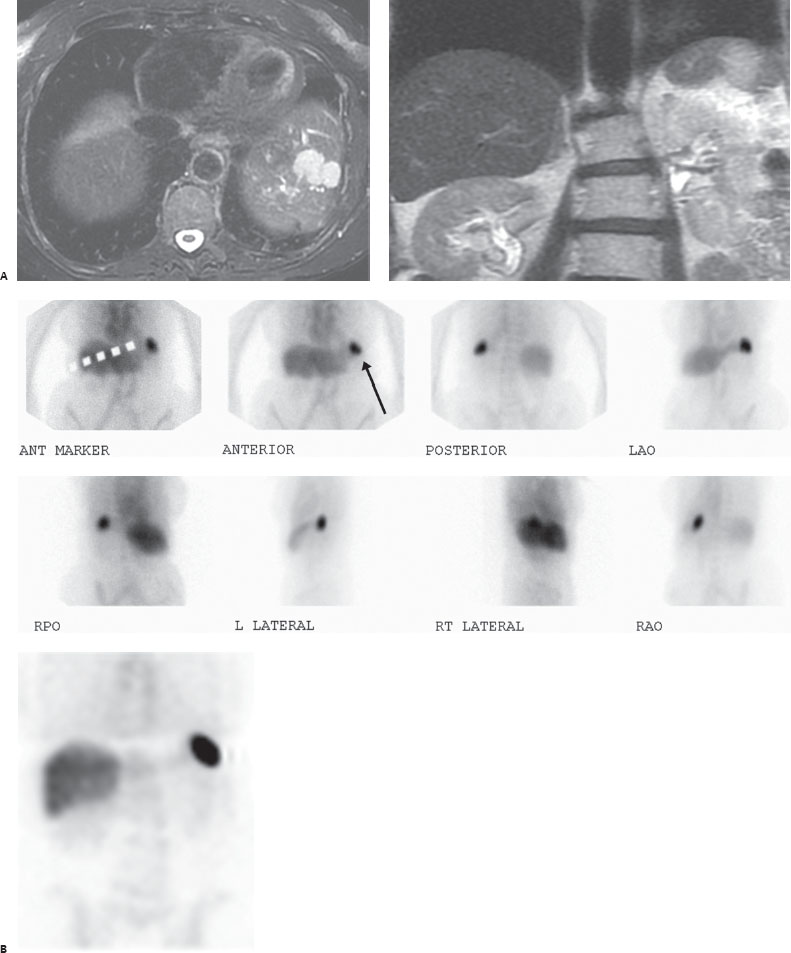
A 67-year-old woman with an incidental mass detected in the left lobe of the liver on MRI. There is a remote history of a motor vehicle accident that resulted in an emergency splenectomy.

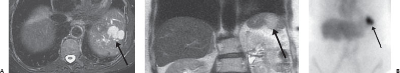
(A) Axial T2 fast spin echo MRI and coronal T2 single shot fast spin echo weighted MRI of the upper abdomen demonstrate a hyperintense lesion at the left lateral liver (arrows). (B) Multiple abdominal spot and coronal SPECT images through the abdomen demonstrate mild physiologic uptake in the blood pool, liver, and bone marrow with an intense focus in the left upper quadrant corresponding to the liver lesion (arrow).
 Differential Diagnosis
• Heat-damaged RBC scan demonstrating splenosis: Mild blood pool and hepatic uptake makes a damaged RBC scan the most likely exam. The more intense focal uptake is most likely splenosis, given the history.
• RBC scan (nondamaged) demonstrating a hepatic hemangioma:
Related posts:


Stay updated, free articles. Join our Telegram channel

Full access? Get Clinical Tree


