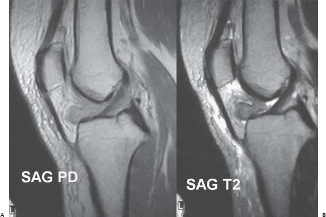
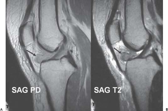
Case 53 A 25-year-old patient presents with loss of knee extension. (A,B) Sagittal proton density- and T2-weighted magnetic resonance (MR) images of the knee show a nodular mass (arrow) between the anterior aspect of the lateral femoral condyle and tibia within the anterior intercondylar notch. The lesion, which has intermediate to low signal intensity, abuts the anterior cruciate ligament (ACL).
 Clinical Presentation
 Imaging Findings
Stay updated, free articles. Join our Telegram channel

Full access? Get Clinical Tree


