Case 53
 Clinical Presentation
A 46-year-old man with vague backache.
 Imaging Findings
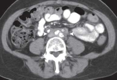
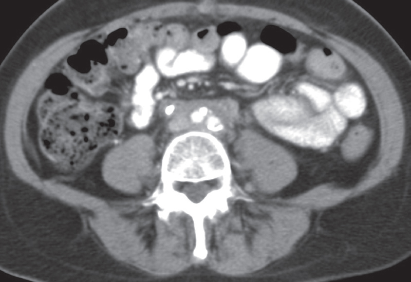
Contrast-enhanced computed tomography (CT) image at the level of the middle abdomen shows a retroperitoneal soft-tissue mass (asterisk) surrounding the bifurcation of the aorta and the inferior vena cava (IVC). The margins are smooth, and the mass does not extend behind the vessels. The ureters (arrowheads) are intimately related to the mass.
 Differential Diagnosis
• Retroperitoneal fibrosis (RPF): Medium-attenuation retroperitoneal soft tissue with smooth margins that is anterior and lateral to the aorta and IVC, and obstructs and displaces the ureters medially is characteristic.
• Retroperitoneal lymphadenopathy:
Stay updated, free articles. Join our Telegram channel

Full access? Get Clinical Tree


