Case 56
Case History
A 73-year-old woman presents with a palpable left breast mass.
Physical Examination
• left breast: palpable mass at the 5:00 position
• right breast: normal exam
Mammogram
Mass (Fig. 56–1)
• margin: indistinct
• shape: irregular
• density: equal
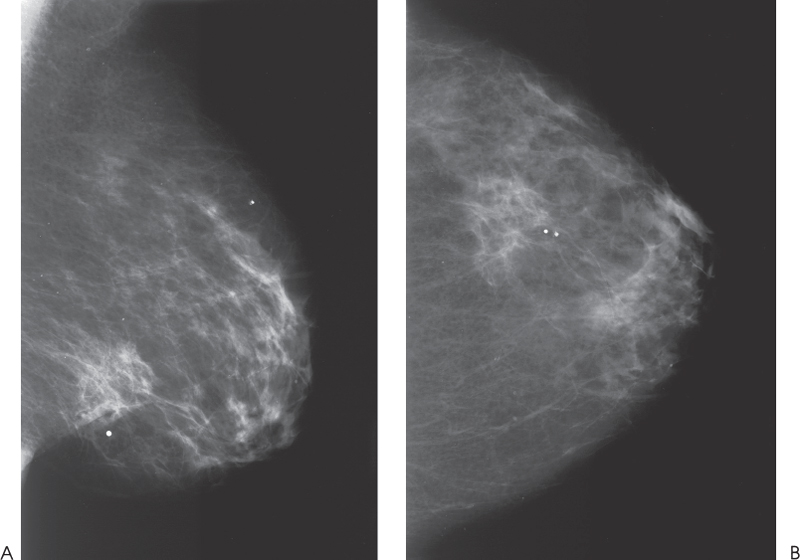
Figure 56–1. An ill-defined mass is in the lower outer quadrant. This mass is associated with skin retraction. This mass corresponds to the palpable lump. (A). Left MLO mammogram. (B). Left CC mammogram.
Ultrasound
Frequency
• 7 MHz
Mass
• margin: spiculation/architectural distortion
• echogenicity: heterogeneous (mixed)
• retrotumoral acoustic appearance: severe shadowing, mass partially obscured


Stay updated, free articles. Join our Telegram channel

Full access? Get Clinical Tree


