 Clinical Presentation
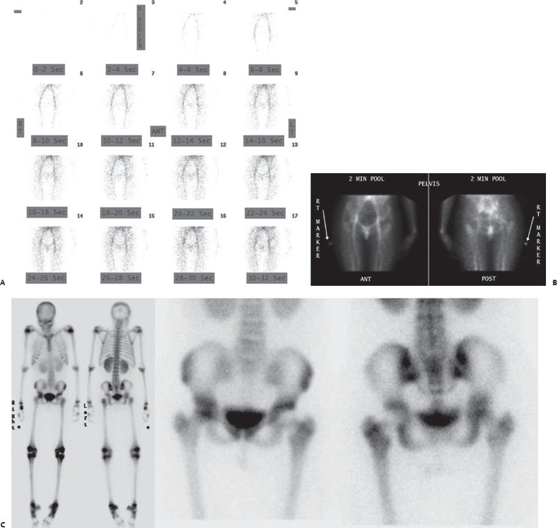
A 15-year-old girl with acute left hip pain.

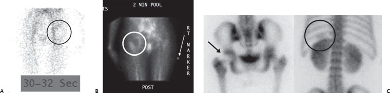
(A) Anterior pelvic angiographic images from a three-phase Tc99m-HDP bone scan show a subtle decrease in blood flow to the left hip (circle). (B) Anterior and posterior pelvic blood pool images reveal an asymmetric region of decreased activity in the left hip (circle). (C) Standard delayed whole-body and pelvic spot images demonstrate a concordant region of decreased uptake involving the left femoral head (arrow). Very subtle splenic soft-tissue uptake is also seen above the left kidney (circle). Symmetric tracer uptake is seen at the epiphyses, which is a normal finding in a child. Focal tracer activity in the right wrist is from injection site artifact.
 Differential Diagnosis
• Acute avascular necrosis (AVN): Acute AVN will present with decreased activity on early and delayed phases of the bone scan.
• Septic arthritis: Typically, blood flow and pool will be increased because of hyperemia; however, increased pressure in the hip joint may cause decreased uptake.
• Tumor:
Related posts:


Stay updated, free articles. Join our Telegram channel

Full access? Get Clinical Tree


