Case 57
Case History
A 59-year-old woman presents with a new density identified by screening mammography.
Physical Examination
• normal exam
Mammogram
Mass (Fig. 57–1)
• margin: indistinct
• shape: irregular
• density: equal
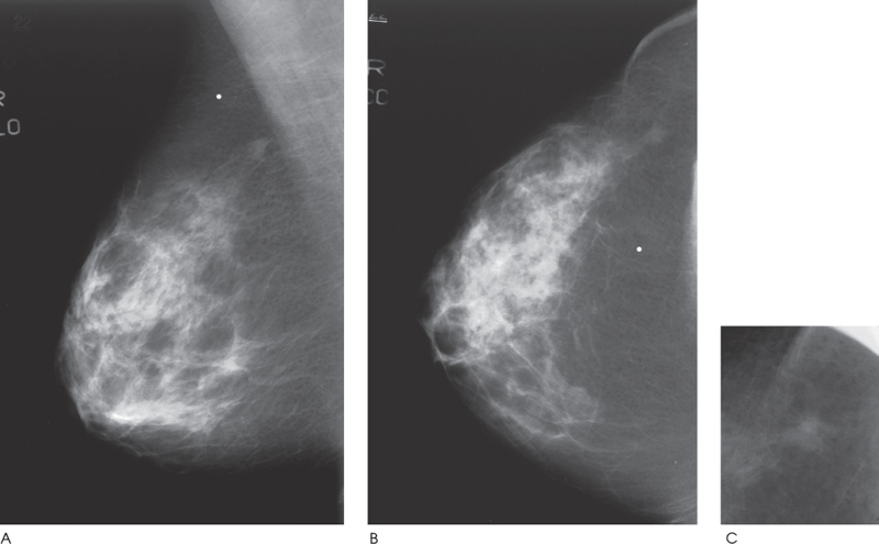
Figure 57–1. There is a small irregular mass in the right upper outer breast. Radiopaque marker (dot) labels a skin mole. (A). Right MLO mammogram. (B). Right CC mammogram. (C). Right CC spot compression mammogram.
Ultrasound
Low Frequency
Frequency
• 7 MHz
Mass
• margin: ill defined


Stay updated, free articles. Join our Telegram channel

Full access? Get Clinical Tree


