Case 58
Case History
A 70-year-old woman presents with non-Hodgkin’s lymphoma and a new right breast mammographic mass.
Physical Examination
• breasts: no palpable masses
• bilateral palpable cervical, axillary, and inguinal adenopathy
Mammogram
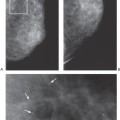
Mass (Fig. 58–1)
• margin: indistinct
• shape: irregular
• density: equal density
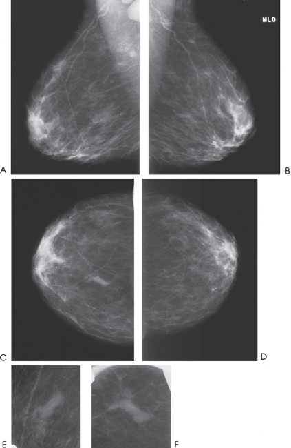
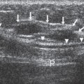
Figure 58–1. In the 12:00 position of the right breast, there is an irregular, ill-defined mass. Large right axillary lymph nodes are also evident. (A). Right MLO mammogram. (B). Left MLO mammogram. (C). Right CC mammogram. (D). Left CC mammogram. (E). Right MLO spot compression mammogram. (F). Right CC spot compression mammogram.
Ultrasound
Frequency
• 13 MHz
Mass
Stay updated, free articles. Join our Telegram channel

Full access? Get Clinical Tree


