Case 58
 Clinical Presentation
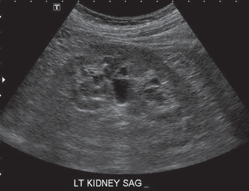
A 58-year-old man who underwent upper abdominal ultrasound for vague abdominal pain.
 Imaging Findings
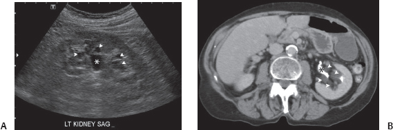
(A) Ultrasound image of the left kidney shows multiple fluid-filled structures (arrowheads) in the renal sinus surrounding the renal pelvis (asterisk), which is mildly prominent. The cystic structures do not appear to communicate with the renal pelvis or one another. (B) Computed tomography (CT) image at the level of the kidneys in the same patient obtained in the excretory phase shows the opacified renal pelvis (black asterisk) to be normal in caliber. It is surrounded by unopacified fluid-containing cystic structures (arrowheads) that correspond to those seen on the ultrasound image in Figure A.
 Differential Diagnosis
• Parapelvic cysts: Multiple noncommunicating cystic-appearing masses, some with branching in the renal sinus, are highly suggestive of parapelvic cysts.
• Bilateral hydronephrosis:
Stay updated, free articles. Join our Telegram channel

Full access? Get Clinical Tree


