Case 59
Case History
A 75-year-old woman presents for a screening mammogram.
Physical Examination
• normal exam
Mammogram
Mass (Fig. 59–1)
• margin: indistinct
• shape: irregular
• density: equal density
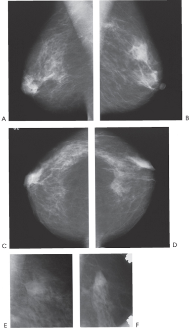
Figure 59–1. In the left upper outer quadrant, there is an ill-defined mass. The mass is not visible on the routine CC view (D) but is identified on the exaggerated CC (F) view. (A). Right MLO mammogram. (B). Left MLO mammogram. (C). Right CC mammogram. (D). Left CC mammogram. (E). Left MLO spot compression mammogram. (F). Left exaggerated CC spot compression mammogram.
Ultrasound
Frequency


Stay updated, free articles. Join our Telegram channel

Full access? Get Clinical Tree


