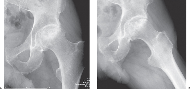
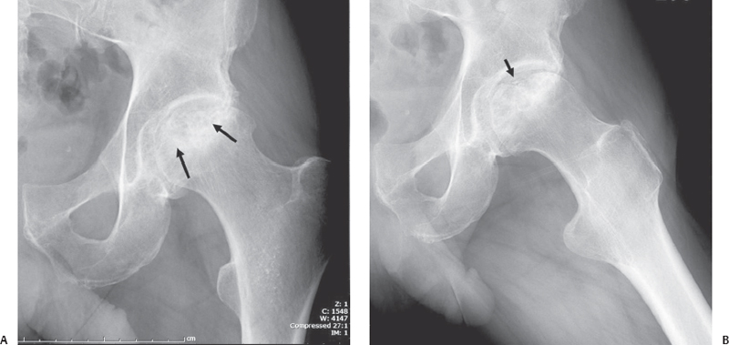
Case 6 The patient is a 52-year-old man with atraumatic pain in his left hip. (A,B) Frontal and lateral radiographs of the left hip show patchy sclerosis (long arrows) involving the femoral head with superior articular collapse (short arrow
 Clinical Presentation
 Imaging Findings
![]()
Stay updated, free articles. Join our Telegram channel

Full access? Get Clinical Tree


