Case 6
Case History
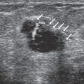
A 48-year-old woman, status post lumpectomy 16 months ago. She now has a small palpable lump in her lumpectomy site. She is initially studied sonographically. Upon discovery of a sonographic nodule, mammographic examination has been performed.
Physical Examination
• left breast: 8 mm lump at the 6:00 position within the scar of her lumpectomy site
• right breast: normal exam
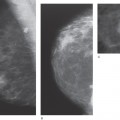
Mammogram
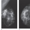
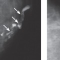
Mass (Fig. 6–1)
• margin: circumscribed
• shape: oval
• density: fat-containing
Stay updated, free articles. Join our Telegram channel

Full access? Get Clinical Tree


