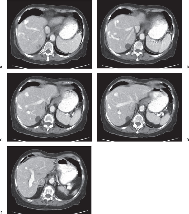
CASE 6 A 36-year-old man presents with mild abdominal pain. Fig. 6.1 (A–E) Axial contrast-enhanced CT images show an irregular linear, hyperattenuating lesion located peripherally within the liver. The lesions appear in close proximity to hepatic veins and portal venous branches and show similar attenuation as the opacified vessels. These findings are indicative of venous malformation. Axial contrast-enhanced computed tomography (CT) images (Fig. 6.1) show two different hyperattenuating, irregularly shaped lesions located peripherally within the hepatic parenchyma, in close proximity to the hepatic veins and the branches of the portal vein. These vessels do not taper peripherally as they are supposed to do but appear otherwise quite prominent. There is early opacification of hepatic veins. Both the lesions present the same attenuation of vessels consistent with vascular perfusion anomalies. Hepatic venous malformation The liver benefits from a dual blood supply: the arterial system accounts for 25% of the total amount, and the portal vein provides the remaining 75%. These two systems are not independent but intimately connected by means of transsinusoidal, transvasal, and transplexal routes. As soon as vascular disorders occur, the dual blood supply undergoes hemodynamic changes concerning both volume and direction of the blood flow. Perfusion alterations represent a relatively common cause of pseudolesions at cross-sectional imaging; thus, radiologists should be aware of these vascular disorders when reading liver CT.
Clinical Presentation
Radiologic Findings
Diagnosis
Differential Diagnosis
Discussion
Background
Clinical Findings
Related posts:


Stay updated, free articles. Join our Telegram channel

Full access? Get Clinical Tree


