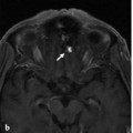
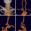
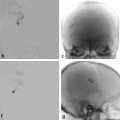
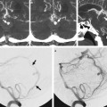
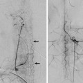
The Dural Ring and the Carotid Cave In a 33-year-old woman with a long-standing history of migraines, an incidental aneurysm was discovered during routine MRI. See ▶ Fig. 6.1. Fig. 6.1 Computed tomographic angiography three-dimensional reconstruction (a) shows a medially oriented aneurysm (double arrows) in the clinoid segment of the ICA, arising below the origin of the ophthalmic artery (arrow). MRI coronal high-resolution T2-weighted images (b,c) show the location of the distal dural ring (DDR on b). The neck of the aneurysm is above the DDR, and the dome is therefore exposed to the subarachnoid space (arrowheads). Left ICA angiogram in oblique view before treatment (d) shows the clinoid segment medially oriented aneurysm before treatment. Plain radiography in oblique view (e) after deployment of two telescoping flow diverter stents demonstrates overlapping of the stents at the neck of the aneurysm. The left ICA angiogram after treatment in arterial (f) and venous (g) phases shows contrast stasis within the aneurysm sac, reflecting a satisfactory flow effect. Unruptured aneurysms of the superior hypophyseal artery above the distal dural ring. The anatomy surrounding the area of the anterior clinoid process (ACP) and the so-called paraclinoid internal carotid artery (ICA) region is complex. The ACP connects medially to the planum sphenoidale via the roof of the optic canal and to the sphenoid body via the optic strut. The optic strut separates the optic canal (medially) from the superior orbital fissure (laterally). There are three dural folds in this region: the falciform ligament, the distal dural ring, and the proximal dural “ring,” also called the carotid-oculomotor membrane (COM). The falciform ligament is the most cranial of the three dural folds. It lies above the optic nerve just proximal to the optic canal entry point, where the nerves enter into the optic canal. This ligament blends medially with the dura that covers the planum sphenoidale. The falciform ligament is an important landmark at the time of microneurosurgical approach to aneurysms in this region. The proximal dural “ring” or COM marks the transition of the cavernous to the clinoid segment of the ICA. The COM lines the lower margin of the ACP. It separates the ACP from the oculomotor nerve, extending from the oculomotor nerve medially to surround the ICA, forming a distinct ring only on the anterior and lateral margins of the artery. The ring is incomplete on the medial side of the ICA. The distal dural ring (DDR) is formed by dura extending medially from the upper surface of the ACP, posteriorly from the upper surface of the optic strut, laterally from the distal end of the carotid sulcus, and anteriorly from the upper surface of the diaphragma sellae and posterior clinoid process. The carotid collar is a thin dural layer between the COM and DDR that surrounds the clinoid ICA. The carotid collar is loosely attached to the ICA except at the DDR, where it blends into a continuous dural layer. The DDR and COM join together to form a single layer at the posterior tip of the ACP, which will blend with the diaphragma sellae. The clinoid segment of the ICA is the portion of the artery located between the proximal and distal dural ring. The DDR is the anatomical landmark that separates the extradural from the intradural ICA, or the clinoid segment from the ophthalmic segment of the ICA. The DDR is firmly attached to the lateral wall of the ICA, but medially, the dura turns downward before attaching to the ICA wall and creating a focal recess. This recess between the DDR and medial ICA wall was called the “carotid cave” by Kobayashi and colleagues in 1989. The importance of this recess lies in the fact that the arachnoid can push through to create a subarachnoid space below the level of the DDR. These recesses can be present in ~75% of individuals, according to anatomic studies in cadavers.
6.1 Case Description
6.1.1 Clinical Presentation
6.1.2 Radiologic Studies

6.1.3 Diagnosis
6.2 Anatomy
Stay updated, free articles. Join our Telegram channel

Full access? Get Clinical Tree


