 Clinical Presentation
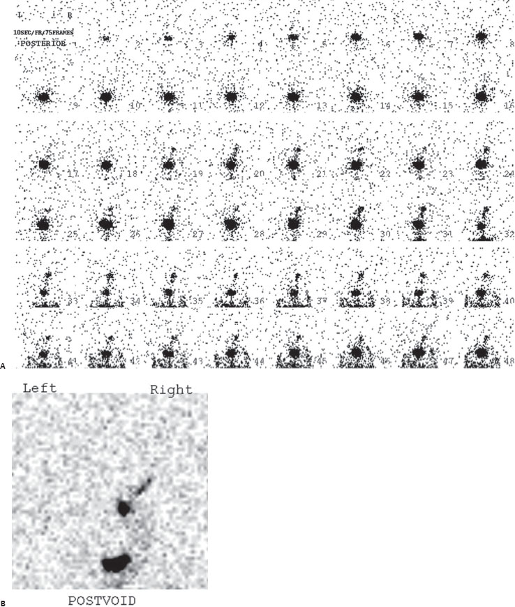
A 1-year-old girl presents after recent urinary tract infection.

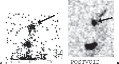
(A) Direct radionuclide cystography was performed with 0.5 mCi of Tc99m-pertechnetate. Dynamic posterior images reveal abnormal radiotracer appearing above the bladder on the right (arrow), increasing during the voiding phase. (B) A static posterior postvoid image demonstrates retention of radiotracer above the bladder on the right (arrow).
 Differential Diagnosis
• Vesicoureteral reflux (VUR): Activity should not be seen above the level of the bladder and if present indicates reflux.
• Urinary contamination artifact: This is commonly seen but appears below the bladder, from leakage around the bladder catheter.
• Urinary leak: This is much less likely and would appear outside the expected location of the bladder and collecting system.
 Essential Facts
• VUR is seen in 35 to 40% of children with urinary tract infection.
Related posts:


Stay updated, free articles. Join our Telegram channel

Full access? Get Clinical Tree


