 Clinical Presentation
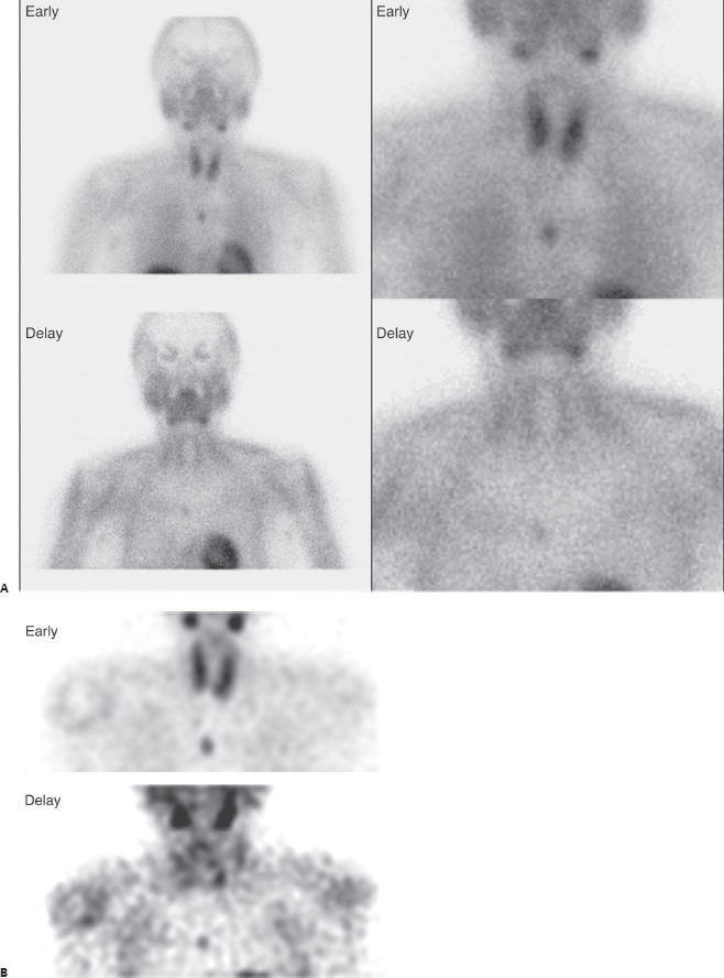
A 52-year-old man with hypercalcemia. Images were acquired 20 minutes and 3 hours after injection.

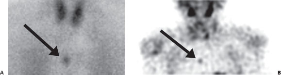
(A) Early planar images demonstrate physiologic radiotracer uptake in salivary, lacrimal, thyroid, and myocardial tissue, making Tc99m-MIBI or -TETRO the most likely agent (parathyroid scan). An abnormal mediastinal focus is seen on early images (arrow); it persists mildly on delayed images. Normal thyroid activity washes out on the delayed planar images. (B) Early and delayed selected coronal SPECT slices demonstrate the abnormal mediastinal focus (arrow). Again, the thyroid is largely washed out on delay.
 Differential Diagnosis
• Ectopic parathyroid adenoma: An abnormal mediastinal focus is most likely this diagnosis, particularly given the history.
• Mediastinal tumor: MIBI and TETRO are nonspecific tracers, and a number of metastatic and primary lesions, including lymphoma, could have this appearance.
• Pulmonary infection/inflammation: Again, these are nonspecific tracers, and an active process could have focal activity.
Related posts:


Stay updated, free articles. Join our Telegram channel

Full access? Get Clinical Tree


