Case 61
Case History
A 57-year-old woman presents with a new left breast lump.
Physical Examination
• left breast: two palpable lumps at the 1:00 position; skin is thickened, red, and erythematous
• right breast: normal exam
Mammogram
Mass (Fig. 61–1)
• margin: indistinct
• shape: irregular
• density: high density
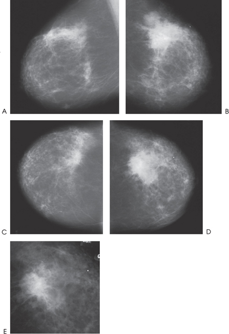
Figure 61–1. In the left upper outer breast there is an area of increased density associated with a dominant ill-defined irregular mass. (A). Right MLO mammogram. (B). Left MLO mammogram. (C). Right CC mammogram. (D). Left CC mammogram. (E). Left CC spot compression mammogram.
Ultrasound
Frequency
• 10 MHz
Mass
• margin: ill defined
• echogenicity: hypoechoic
• retrotumoral acoustic appearance: posterior shadowing distal to mass


Stay updated, free articles. Join our Telegram channel

Full access? Get Clinical Tree


