Case 64
 Clinical Presentation
A 34-year-old woman with recurrent pelvic pain.
 Imaging Findings
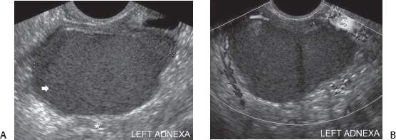
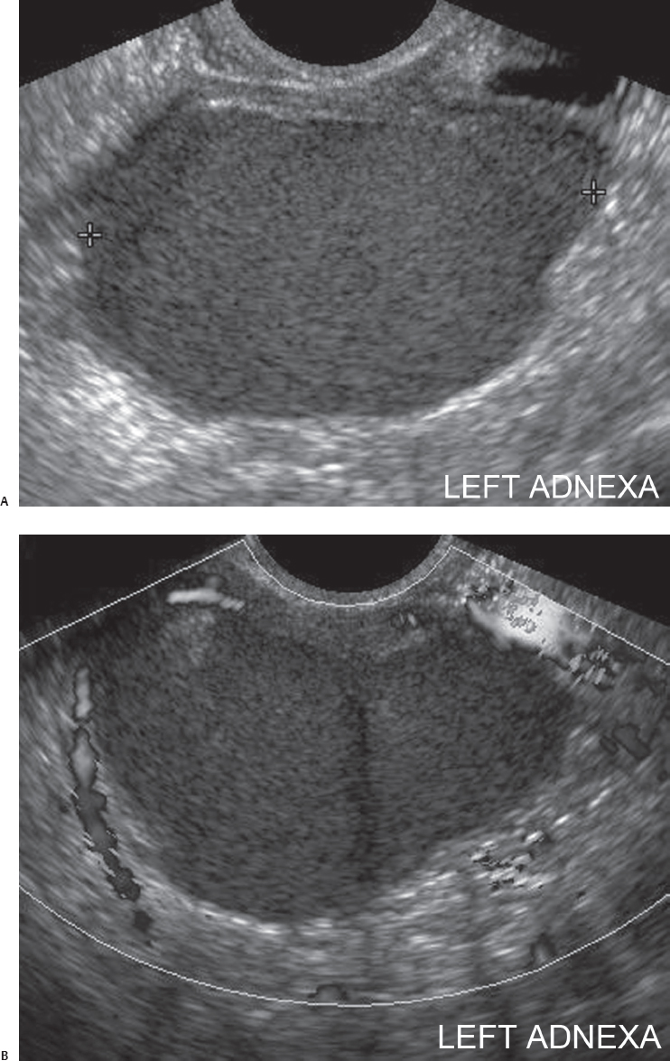
(A) Transvaginal sonographic image of the left adnexa shows an adnexal mass (arrow). It is hypoechoic with uniform low-level echoes. There is distal acoustic enhancement (asterisk). The wall is well formed. (B) Power flow ultrasound image of the mass in Figure A shows absence of flow within the lesion.
 Differential Diagnosis
• Endometrioma: A cystic lesion with low-grade internal echoes is characteristic.
• Hemorrhagic ovarian follicle:
Stay updated, free articles. Join our Telegram channel

Full access? Get Clinical Tree


