 Clinical Presentation
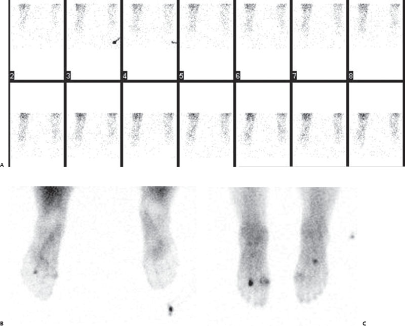
A 72-year-old woman with a history of diabetes and a nonhealing wound in the plantar aspect of the left foot.

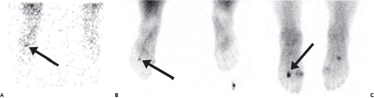
(A) Plantar flow images reveal mild, diffuse increase in activity involving the left foot. Slightly increased focal activity is seen in the region of the third metatarsal (arrow). (B) Plantar blood pool images reveal focal increase in activity in the region of the left third metatarsal (arrow). (C) Plantar delayed images reveal intense focal uptake at the left third distal metatarsal (arrow). Less pronounced uptake is seen at the left first metatarsophalangeal joint and in the right midfoot (regions where uptake was not significantly increased on the earlier phases and where it is likely related to degenerative changes).
 Differential Diagnosis
A three-phase positive bone scan can be seen with fracture (stress or completed), infection, or even tumor.
• Osteomyelitis: Focal three-phase positive uptake is the diagnosis of exclusion, given the history.
Related posts:


Stay updated, free articles. Join our Telegram channel

Full access? Get Clinical Tree


