Case 67
 Clinical Presentation
A 39-year-old woman referred from the emergency department to investigate vague suprapubic pain.
 Imaging Findings
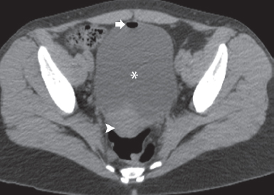
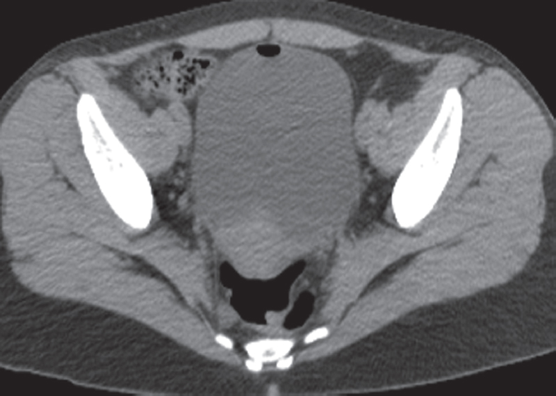
Non–contrast-enhanced computed tomography of the pelvis in the region of the urinary bladder. The urinary bladder is filled with clear urine (asterisk). There is an air bubble (arrow) in the nondependent portion of the urinary bladder. The urinary bladder wall is normal in thickness. There is no stranding in the pelvic fat. The uterus (arrowhead) appears normal.
 Differential Diagnosis
• Recent instrumentation: Air enters the urinary bladder most commonly as a consequence of catheterization. The absence of any other abnormality favors that diagnosis. This patient was catheterized in the emergency room to obtain a clean urine sample.
• Fistula with the bowel: Gas in the urinary bladder lumen in the absence of recent instrumentation is diagnostic of a fistula with the bowel. Depending on the site of the fistula and the communicating portion of the bowel, a localizing sign such as focal bladder wall thickening, bowel wall thickening, or a mass and fat stranding would be expected.
Stay updated, free articles. Join our Telegram channel

Full access? Get Clinical Tree


