 Clinical Presentation
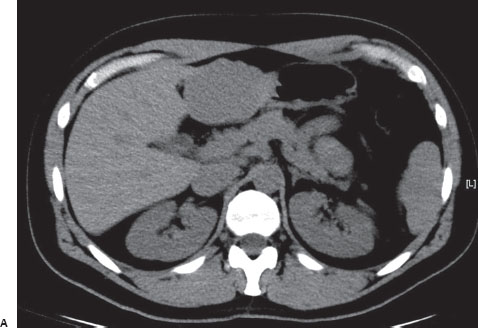
A 34-year-old woman with left upper quadrant pain.
Further Work-up
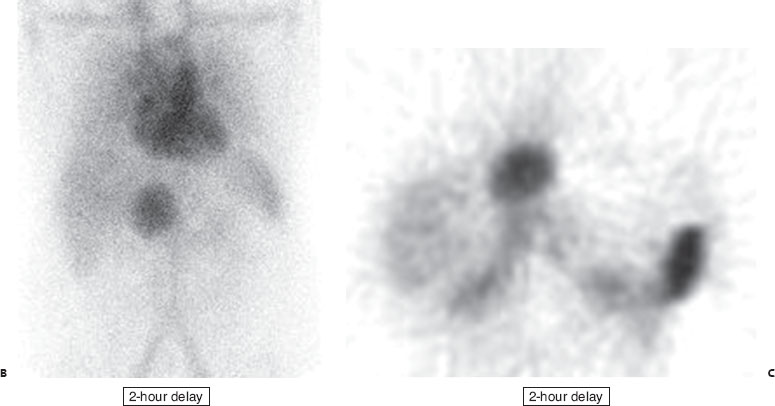

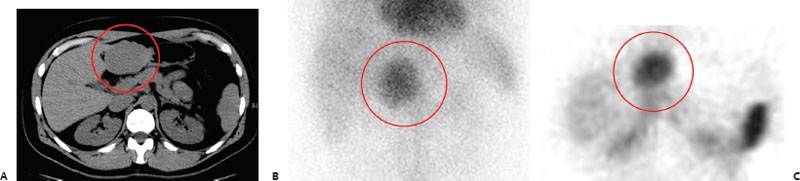
(A) Noncontrast CT demonstrates a hypodense mass in the medial segment (segment 3) of the left lobe of the liver (circle). (B,C) Two-hour–delayed anterior planar and axial SPECT images of the abdomen from a Tc99m-RBC scan demonstrate a corresponding focus of increased activity (circles).
 Differential Diagnosis
• Cavernous hemangioma: Focally increased RBC accumulation on delayed blood pool imaging is virtually pathognomonic for this entity.
• Angiosarcoma: This can reportedly have the same appearance but is exceedingly rare.
• Focal nodular hyperplasia: This is the second most common benign tumor of the liver but is cold on an RBC scan (often hot on SCOL and HIDA).
 Essential Facts
Related posts:


Stay updated, free articles. Join our Telegram channel

Full access? Get Clinical Tree


