Diagnosis: Bennett fracture-dislocation of the first metacarpal base
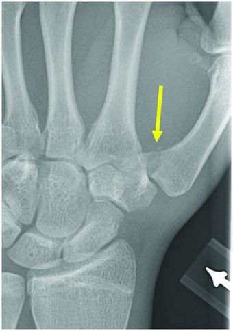
PA radiograph of the hand demonstrates an intra-articular fracture through the thumb metacarpal base (arrow), with mild radial subluxation of the dominant radial metacarpal fracture fragment.
Discussion
Overview and mechanism of Bennett fracture
A Bennett fracture is an intra-articular fracture through the ulnar articular margin of the volar beak of the base (arrow) of the thumb metacarpal, with varying degrees of associated subluxation or dislocation of the radial metacarpal fragment by traction from the abductor pollicis longus tendon. The smaller ulnar fragment remains in place due to anchoring by the attached anterior oblique ligament.
The mechanism most commonly involves axial compression on a partially flexed thumb.
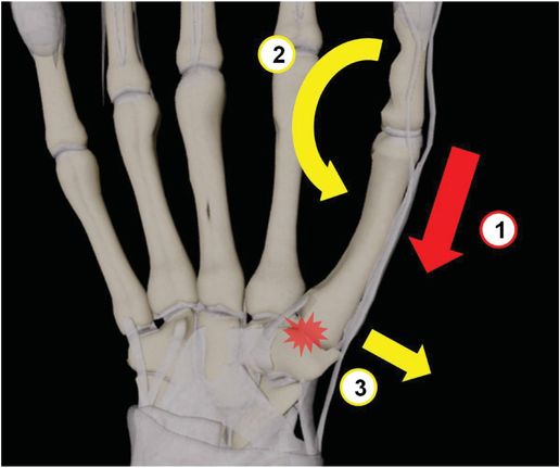
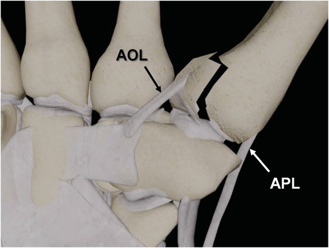
In the Bennett fracture, the small fracture fragment at the ulnar base of the first metacarpal is held in place by the anterior oblique ligament (AOL), and the radial component of the first metacarpal subluxes laterally under tension from the abductor pollicis longus tendon (APL).
Imaging of Bennett fracture
Dedicated PA radiographs of the thumb base with the forearm in slight pronation are often required to adequately characterize the fracture fragment size.
CT can be used to better characterize the degree of articular involvement in cases with ambiguous plain radiograph findings.
Treatment of Bennett fracture
Closed reduction can be attempted if there is <3 mm of fragment displacement, although percutaneous pin fixation may be ultimately required.
Open reduction and internal fixation (ORIF) is often required with fracture fragment displacement >3 mm. Other indications for ORIF include significant impaction of the central articular surface seen on CT, clinical findings of neurovascular injury, or failure of closed reduction.
Clinical synopsis
The patient underwent closed reduction with percutaneous pinning.
Differential diagnosis of Bennett fracture
Rolando fracture
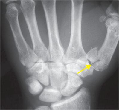
Rolando fracture is a ≥3-part fracture of the thumb metacarpal base, which can be described as Y-shaped, T-shaped, or comminuted. These injuries typically require open reduction and internal fixation, with generally worse post-treatment functional outcomes in contrast to Bennett fractures, which are non-comminuted (hence the mnemonic “Bennett is better”). Radiograph of the hand demonstrates a three-part, intra-articular (arrow) fracture of the thumb metacarpal base with a Y-shaped configuration.
Related posts:
 12 68-year-old man with left lower quadrant pain and hypotension
 32 32-year-old male complaining of chest pain after upper endoscopy
 28 30-year-old male presented with a palpable left testicular mass
 29 19-year-old male presented with acute onset right scrotal pain
 53 42-year-old female presenting with fever and back pain
 66 21-year-old male with quadriplegia after diving into a shallow pond. The patient struck his head against an embankment, with his head flexed, chin against chest
Stay updated, free articles. Join our Telegram channel

Full access? Get Clinical Tree


