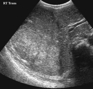
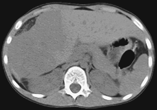
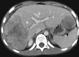
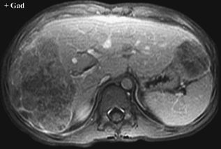
CASE 69 An 8-year-old boy presents with abdominal pain. Figure 69A Figure 69B Figure 69C Figure 69D A large echogenic mass, poorly marginated, is noted in the right lobe of the liver on ultrasonography (Fig. 69A). A heterogeneous mass without calcification is shown on nonenhanced CT (Fig. 69B). Following contrast administration, peripheral enhancement of two lesions, one in the right lobe and one in the left lobe (Fig. 69C), is noted. The corresponding delayed T1-weighted fat suppressed MRI postgadolinium reveals heterogeneous signal of the masses, which are not hypervascular (Fig. 69D). Hepatoblastoma Hepatoblastoma is the most common pediatric hepatic malignancy (>50%) and is most prevalent in children <3 years of age. Male to female ratio is 1.2–3.3:1. An increased incidence has been reported in Beckwith-Wiedemann syndrome, hemihypertrophy, familial adenomatous polypi, glycogen storage disease, Wilms’ tumor, Gardner’s syndrome, and fetal alcohol syndrome. Low birth weight is associated with a higher risk for hepatoblastoma. Cytogenetic abnormalities have been noted with mainly abnormalities of chromosome 20, and a recurring aberration der (4) t (1q;4q). Alteration in the adenomatous polyposis coli (APC) gene and the b-catenin pathway are implicated in the pathogenesis of hepatoblastoma. Changes in the expression of H19 and IGF2 genes have also been found.
Clinical Presentation
Radiologic Findings
Diagnosis
Differential Diagnosis
Discussion
Background
Etiology
Molecular Biology
Clinical Findings
Related posts:


Stay updated, free articles. Join our Telegram channel

Full access? Get Clinical Tree


