7 Image Processing
Introduction
Image processing and display are critical components in the imaging workflow chain. They have tremendous diagnostic utility for disease detectability and interpretation. In addition, the combination of information from two or more modalities can increase both sensitivity and specificity compared to a single exam. This chapter presents basic image processing concepts that are used in medical imaging.
7.1 Case 1: Filtering and Edge Enhancement
7.1.1 Background
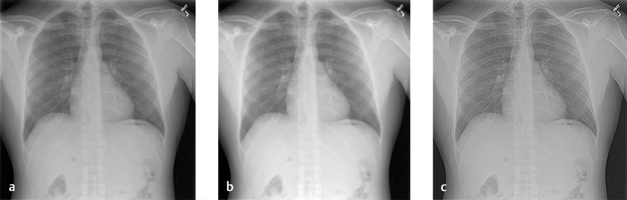
Presentation of an anteroposterior (AP) chest radiograph.
7.1.2 Findings
Presentation of post image filtering by convolution with kernels designed to extract features of different frequencies.
Kernels are commonly used to lower noise or enhance edge information.
7.1.3 Discussion
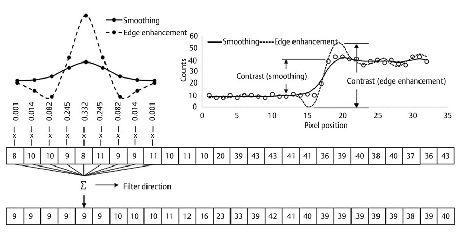
Filtering is one of the most basic imaging processing steps to enhance image contrast. It can be applied either in frequency space through use of the Fourier transform or in image space through use of the convolution process. 1 In image space, filtering involves construction of a kernel that is moved across the image. All pixels within the kernel are averaged and that average is placed in a new image. A common kernel is a Gaussian function, which is a low-pass operation that reduces noise (e.g., quantum mottle) and improves visibility of low-contrast features. High-contrast features can be enhanced by adding negative lobes to the kernel. This process improves high-contrast resolution but also increases noise since both of these features are high-frequency components of an image. Kernels are normalized to preserve the scale of the original image. The convolution process is described in Fig. 7‑1 1 where discrete kernel values are multiplied by the pixel values that fall underneath the function and the sum of these products is placed in a new image. The kernel is then shifted and the process is repeated. Fig. 7‑2 demonstrates the change in contrast of a chest radiograph following Gaussian smoothing and Gaussian–Laplacian edge enhancement.
7.1.4 Resolution
Convolution using kernels designed to lower noise or enhance edge information alters contrast and can improve the detectability of anatomical features such as soft-tissue masses or bone fractures.
7.2 Case 2: Maximum Intensity Projection
7.2.1 Background
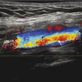
Whole-body [18F] fluorodeoxyglucose ([18F] FDG) positron emission tomography (PET) of a patient with extensive disease.
The image volume is processed with a ray-tracing technique called maximum intensity projection (MIP), to highlight hyper-metabolic activity throughout the volume (Fig. 7‑3).
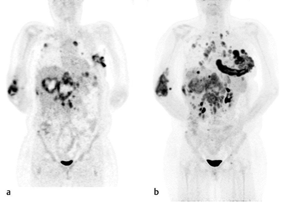
Fig. 7.3 (a) Coronal slice of a [18F] FDG whole-body PET/CT showing extensive disease. (b) Maximum intensity projection processed volume in the coronal orientation along the anterior-posterior direction. Note the visualization of lesions at deeper slices that are out of plane in (a).
7.2.2 Findings
Compared to a standard coronal slice, the MIP image permits visualization at depth within a 3D volume on a 2D display.
The MIP volume can be rotated to improve visualization of hot lesions that lie along the same ray path but in different planes.
Related posts:

Stay updated, free articles. Join our Telegram channel

Full access? Get Clinical Tree


