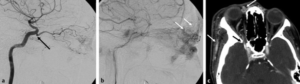
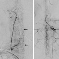
The Dorsal and Ventral Ophthalmic Arteries A 22-year-old man imaged before retreatment of a facial arteriovenous malformation (AVM). See ▶ Fig. 7.1. Fig. 7.1 Left ICA angiogram in lateral view in early (a) and late (b) phases shows that the ophthalmic artery originates at the junction between the horizontal cavernous and the clinoid segment (arrow), within the region of the ILT, and supplies the facial compartment of a diffuse AVM (white arrows in b). Contrast-enhanced computed tomography (c) demonstrates the abnormal vessels over the roof of the nose and along the right side of the face (white arrows). The ophthalmic artery enters the orbit through the superior orbital fissure (black arrow in c). Dorsal ophthalmic artery supplying the orbital component of a facial AVM. The orbit contains structures derived from different embryonic layers: the neuroectoderm (optic nerve [ON], retina, ciliary bodies), the paraxial mesoderm (musculoaponeurotic system, bone, cartilage), and the surface ectoderm (lens, lacrimal gland, eyelids). In humans, there are three arterial systems that potentially participate in the supply to the orbit: the primitive ventral ophthalmic artery (PVOA), the dorsal ophthalmic artery (DOA), and the orbital artery, a branch of the stapedial artery that constitutes the future middle meningeal artery (see Case ▶ 3). As such, the final appearance of the ophthalmic artery (OA) and the blood supply to the orbit in the adult are the result of multiple regressions and anastomoses that occur early in embryonic life. The PVOA arises from what will become the A1 segment of the anterior cerebral artery (ACA). This artery supplies the structures derived from the neuroectoderm and will follow the path of the ON, which is an “orbital extension” of the central nervous system. This artery enters the orbit through the optic canal, inferior and medial to the ON, and gives rise to the central retinal artery and the nasal ciliary artery. The DOA arises from the cavernous (or fifth) segment of the internal carotid artery (ICA), enters the orbit through the superior orbital fissure, and courses inferior and lateral to the ON. This artery gives off the temporal ciliary artery. The orbital artery (branch of the future middle meningeal artery) enters the orbit via the superior orbital fissure. It then divides into a medial ethmoidonasal branch and a lateral lacrimal branch. Both vessels will follow the various rami of the ophthalmic division of the trigeminal nerve (V1), providing supply to the musculoaponeurotic system and the eyelids. According to Lasjaunias et al, in the embryo, the PVOA and the DOA anastomose in the orbit near the ON. In a process that is not predictable, the proximal part of the PVOA and DOA regresses and a new anastomosis develops between the supraclinoid ICA and the PVOA, forming the origin of the future OA. The remnant of the DOA is part of the inferolateral trunk (ILT). At the 20-mm stage, the ethmoidonasal (medial) branch of the orbital artery (branch of the future middle meningeal artery) crosses from lateral to medial over the ON to anastomose with what will become the orbital segment of the OA. At this stage, a true ON anastomotic ring has formed. The OA starts to assimilate the orbital artery, and at the 40-mm stage of the embryo, this process is completed and the configuration of the “adult” OA becomes apparent.
7.1 Case Description
7.1.1 Clinical Presentation
7.1.2 Radiologic Studies
7.1.3 Diagnosis
7.2 Embryology and Anatomy
Related posts:


Stay updated, free articles. Join our Telegram channel

Full access? Get Clinical Tree


