 Clinical Presentation
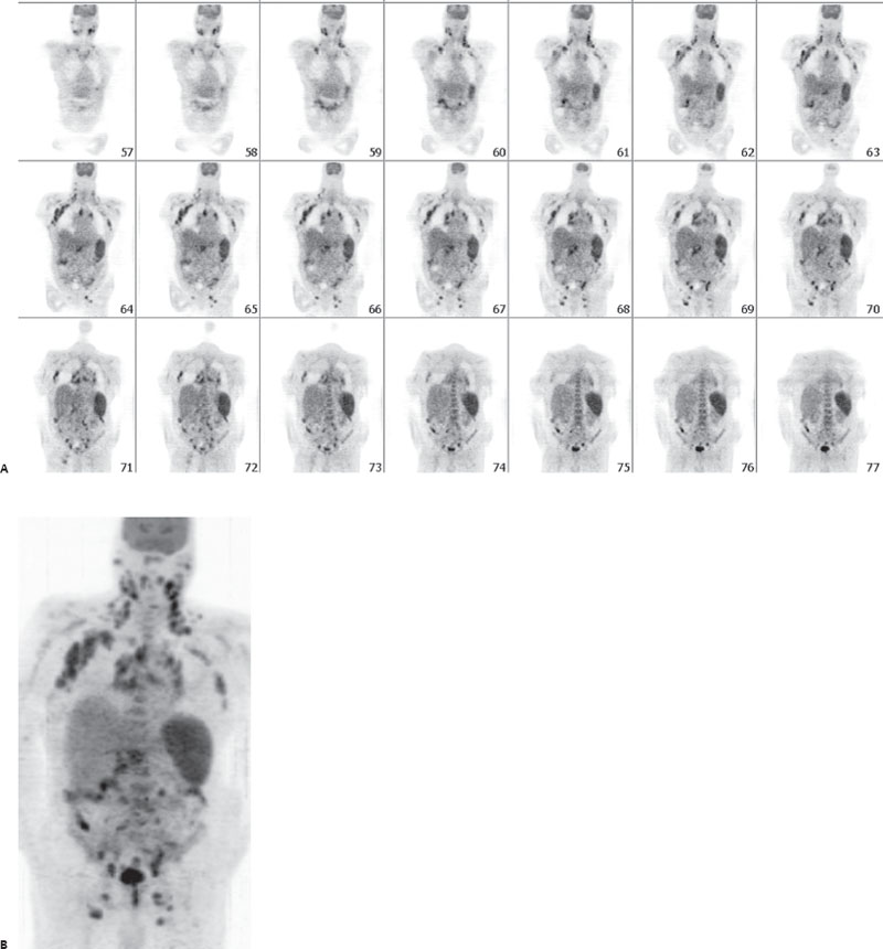
An 84-year-old man with weight loss.

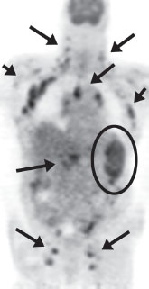
Coronal and anterior MIP images from a whole-body exam demonstrate normal brain gray matter, making this most likely an FDG-PET. Multiple abnormal foci are seen corresponding to lymph node stations in the neck, chest, abdomen, and pelvis (arrows), as well as diffuse abnormal splenic uptake (circle).
 Differential Diagnosis
• FDG-PET demonstrating lymphoma: Widespread symmetric hypermetabolic lymph nodes as well as diffuse splenic involvement make this the most likely etiology.
• FDG-PET demonstrating sarcoidosis: This is more commonly confined to the chest but can appear more diffusely and symmetrically, as in this case. It would be a more likely consideration in a younger female.
• FDG-PET demonstrating diffuse metastatic disease from another primary neoplasm: This can occasionally be this diffuse, such as with melanoma, but the symmetric involvement and diffuse splenic involvement would be less likely.
Related posts:


Stay updated, free articles. Join our Telegram channel

Full access? Get Clinical Tree


