 Clinical Presentation
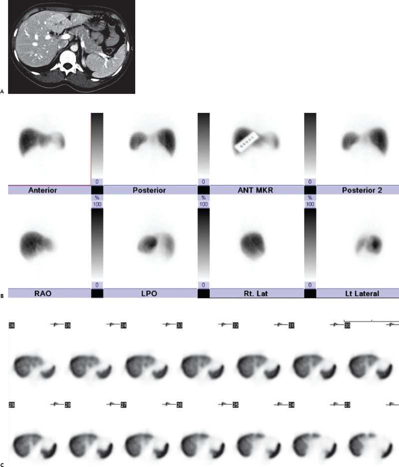
A 32-year-old woman with a left lobe liver lesion seen on CT, performed to evaluate abdominal pain.

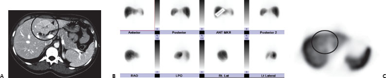
(A) Axial CT image reveals an enhancing mass in the left lobe of the liver with a central scar (circle). (B) Planar images from a Tc99m-SCOL scan reveal homogeneous tracer uptake in the liver and spleen. No colloid shift is seen. No focal abnormalities are seen in the left lobe of the liver. (C) Transverse SPECT images reveal the region corresponding to the CT abnormality has significant Tc99m-SCOL accumulation, similar although slightly less compared to the remainder of the liver (circle).
 Differential Diagnosis
• Focal nodular hyperplasia (FNH): This contains Kupffer cells and thus usually demonstrates uptake similar to or greater than that in surrounding liver. In a young, asymptomatic patient, this is the most likely diagnosis.
• Regenerating nodule: This also contains Kupffer cells and thus shows normal tracer uptake. It is associated with cirrhosis and would not have a central scar on CT.
• Hemangioma:


Stay updated, free articles. Join our Telegram channel

Full access? Get Clinical Tree


