 Clinical Presentation
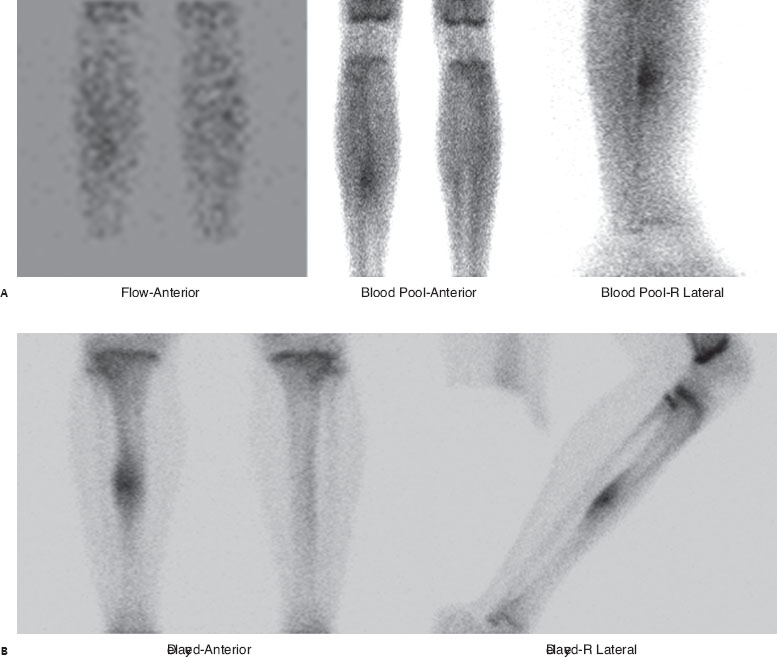
A 16-year-old boy with right leg pain that is most pronounced at night.

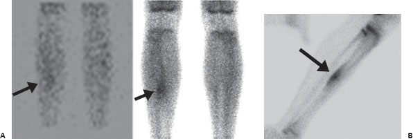
(A) Anterior and right lateral images of the lower extremities from a Tc99m-MDP three-phase bone scan demonstrate focal increase in flow and blood pool activity in the right mid tibia (arrows). (B) The delayed anterior image reveals fusiform focal uptake in the right mid tibia. The lateral view localizes this uptake to the posterior tibial cortex. Centrally, there is a very small focal region of more intense uptake (arrow). Increased uptake at multiple physes is seen, which is appropriate for the patient’s age.
 Differential Diagnosis
• Osteoid osteoma:
Related posts:


Stay updated, free articles. Join our Telegram channel

Full access? Get Clinical Tree


