 Clinical Presentation
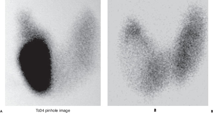
A 56-year-old woman with a palpable right thyroid nodule presents for evaluation. Pertechnetate images were acquired first. Iodine 123 and US images were obtained 2 weeks later.
Further Work-up
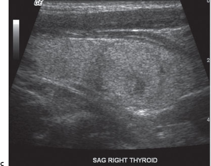

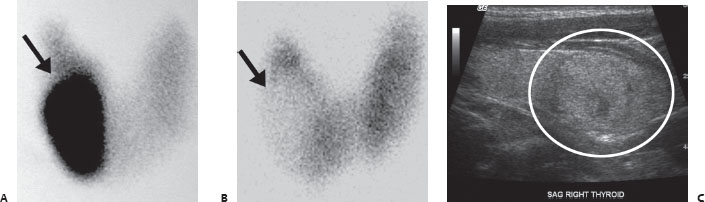
(A) Anterior pinhole image from a pertechnetate (TcO4) thyroid scan reveals focal increase in radiotracer accumulation in a large right lower pole nodule (arrow). Normal homogeneous activity is seen throughout the remainder of the gland. (B) Anterior pinhole image from an iodine 123 thyroid scan reveals focal decrease in activity in the right lower pole nodule (arrow). Normal homogeneous activity is seen in the remainder of the gland. (C) Longitudinal thyroid US image from this patient demonstrates a medium-sized predominantly solid lesion in the right mid region and lower pole (circle).
 Differential Diagnosis
• Discordant nodule indeterminate for malignancy: Nodules that accumulate significant TcO4


Stay updated, free articles. Join our Telegram channel

Full access? Get Clinical Tree


