 Clinical Presentation
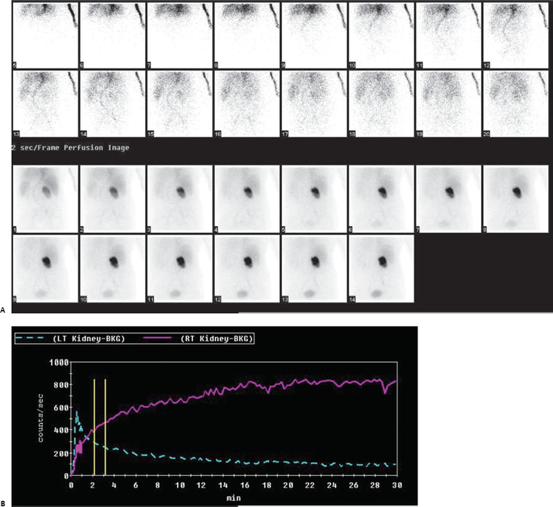
A 60-year-old woman with elevated creatinine and oliguria.

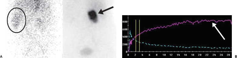
(A) Posterior images from a Tc99m-MAG3 renal scan. Flow images reveal decreased tracer activity in the right kidney. There is apparent flow to the left kidney; however, this represents splenic activity (circle). The patient had a left nephrectomy. Dynamic images reveal abnormal progressive tracer parenchymal extraction by the right kidney (arrow) but only minimal evidence of urinary excretion. (B) Curves generated from a region of interest around the entire kidney show a continually rising renogram on the right (arrow). The left renal curve appears to demonstrate some function because the technologist erroneously drew a region of interest around the spleen.
 Differential Diagnosis
Related posts:


Stay updated, free articles. Join our Telegram channel

Full access? Get Clinical Tree


