The Branches of the Ophthalmic Artery
8.1 Case Description
8.1.1 Clinical Presentation
A 54-year-old man presented with a long-standing pulsatile mass in the left medial periorbital region.
8.1.2 Radiologic Studies
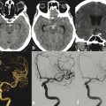
See ▶ Fig. 8.1.

Fig. 8.1 Left ICA angiogram in lateral view (a) shows an AVM nidus in the medial orbit, supplied mainly by the medial palpebral arteries, draining via the angular vein to the common facial vein (white arrow). Superselective microcatheter injection in the OA (b) proximal to the characteristic OA bend shows opacification of the long ciliary arteries (black double arrows) and the choroidal blush, meaning the catheter is proximal to the CRA, which is thus an unsafe position for embolization (see following). Note the anterior falcine artery (black arrowhead), which is a distal branch of the anterior ethmoidal artery. After a more distal catheterization and liquid embolic embolization, the left ICA angiogram in lateral view (c) shows no residual AV shunting and persistent that choroidal blush, indicating that the CRA was preserved.
8.1.3 Diagnosis
Medial orbital arteriovenous malformation (AVM) with main supply derived from the ophthalmic artery (OA).
8.2 Anatomy
The blood supply of the orbit depends mainly on the OA, with minor contributions from the external carotid artery (ECA). The intraorbital segment of the OA gives rise to numerous arterial branches that supply the orbital structures. The branching pattern of the OA shows a high variability in the order and site of origin of the different branches. Conceptually, these branches can be divided into four groups: the ocular group (central retinal and ciliary arteries), the orbital group (lacrimal and muscular arteries), the extraorbital group (ethmoidal, supraorbital, supratrochlear, palpebral, and dorsal nasal arteries), and the dural group (recurrent deep and superficial arteries). Typically, these branches are annexed by the OA and form the “adult” pattern of the OA.
8.2.1 Ocular Group
The ocular group originates from the embryonic internal carotid artery (ICA), via the ventral OA (hyoid branch) and shows little variability in its branching pattern. The order in which the ocular group branches originate is independent of the OA origin but depends on whether the artery courses over (83% of cases) or under (17% of cases) the optic nerve (ON). When the OA courses over the ON, the first branch is the central retinal artery (CRA), followed by a long posterior ciliary artery. When the OA courses under the ON, the first branch is the long lateral posterior ciliary artery and the second is the CRA. The CRA is a terminal branch, with no well-established anastomoses. Therefore, compromising this artery during embolization procedures will invariably result in retinal ischemia and monocular blindness. The long posterior ciliary arteries may arise separately or as part of a musculociliary trunk. They supply the choroid and are responsible for the choroidal blush, which serves as a surrogate marker for the CRA, as the ciliary arteries and the CRA arise closely together from the OA.
8.2.2 Orbital Group and Lacrimal Artery
Stay updated, free articles. Join our Telegram channel

Full access? Get Clinical Tree


