 Clinical Presentation
A 30-year-old man with colorectal cancer (mucinous type) status post resection and chemotherapy presents for restaging. He is also status post bladder reconstruction. Baseline CT (not shown) was normal. No presurgical FDG-PET was performed.

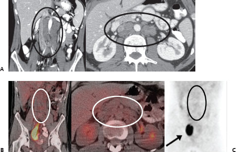
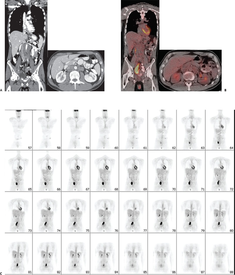
(A) Selected coronal and axial slices from contrast-enhanced CT demonstrate multiple enlarged retroperitoneal lymph nodes (circles), which are new from the previous scans. (B,C) Coronal whole-body FDG-PET images and selected axial and coronal hybrid PET/CT images demonstrate no abnormal FDG uptake to correspond to the enlarged retroperitoneal lymph nodes or elsewhere (circles). Physiologic excretion is noted in the reconstructed bladder in the right pelvis (arrow).
 Differential Diagnosis
• Progression of non–FDG-avid colorectal metastases: Abnormal enlarging lymph nodes on CT in a patient with a known cancer are highly suspicious for metastatic disease, even without FDG uptake. Mucinous cancers are often non–FDG-avid, so this is the diagnosis of exclusion.
• Development of secondary lymphoproliferative neoplasm:


Stay updated, free articles. Join our Telegram channel

Full access? Get Clinical Tree


