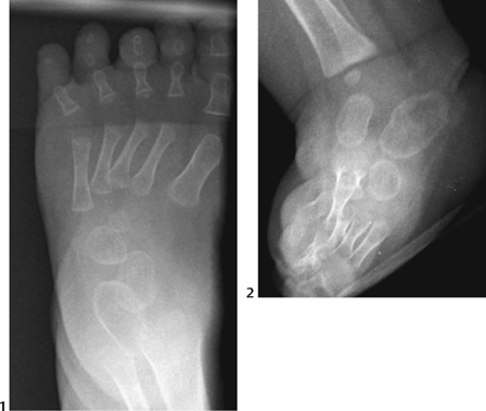
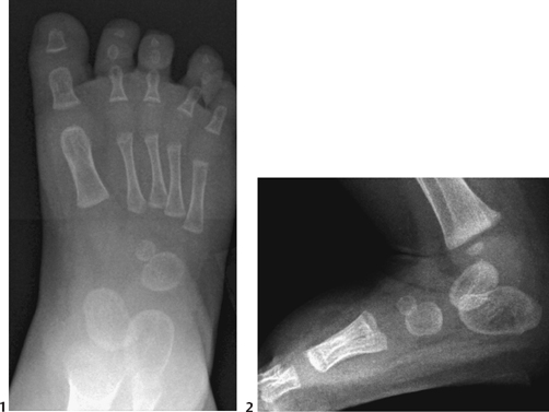
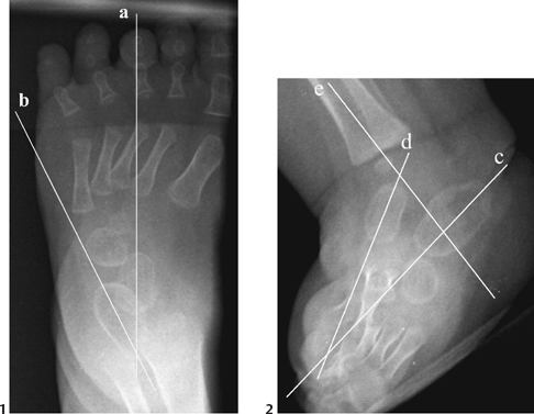
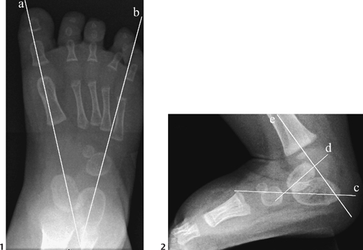
CASE 87 An otherwise healthy newborn male presents with left foot deformity. Figure 87A Figure 87B The long axis of the left talus points lateral to the base of the first metatarsal on the frontal film. There is a horizontal alignment of talus and calcaneus on both frontal and lateral radiographs (Fig. 87A). The calcaneus is in an equinus position. The forefoot is inverted resulting in a stepladder appearance to the metatarsals on the lateral view. The normal right foot is shown for comparison (Fig. 87B). Figure 87C (1) Frontal radiograph of foot demonstrates alignment of the long axis of talus with the base of second metatarsal (line a). Metatarsals are adducted. Cuboid ossification appear shifted medially on calcaneus. Long axis of calcaneus (line b) lies lateral to base of fifth metatarsal with cuboid shifted medially. (2) Lateral foot radiograph demonstrates a stepladder appearance to the metatarsals with the 1st metatarsal most superior. The talus and calcaneus have a more parallel alignment (angle formed by lines d and c). The calcaneus is in equinus position relative to long axis of tibia (angle formed by lines e and c). Left congenital equinovarus or clubfoot with normal right foot for comparison Clubfoot is commonly subdivided into four types: congenital, teratogenic, syndromic, and positional. The congenital variety is the most common, with an incidence of 1 in 1000. This usually isolated deformity is more common in males (2:1) and is bilateral in up to 50% of cases. First-degree relatives are estimated to have a 30x increased incidence compared with the general population. The etiology for the congenital form is uncertain. Defective connective tissues with ligamentous laxity, abnormal types and number of muscle fibers, vascular hypoplasia, and defective anterior horn cells have been reported in pathologic studies. A resulting deformity of the talus is suspected to lead to the altered angular relationships of the foot. The teratogenic type is found in children with underlying disorders such as arthrogryposis. The positional variety is the result of a normally formed foot held in an abnormal intrauterine position as in oligohydramnios. Figure 87D (1) Frontal radiograph demonstrates normal angular relationship of bone structures. Long axis of talus (line a) points to base of first metatarsal. Long axis of calcaneus (line b) points to base of fourth through fifth metatarsals. (2) Lateral radiograph depicts normal angular relationship of bone structures of foot. Normal dorsiflexion of calcaneus, as depicted by angle formed by the long axis of tibia (line e), intersecting long axis of calcaneus (line c). Normal angular relationship of talus (line d) with calcaneus (line c). On clinical examination the infant with clubfoot displays a variable degree of foot inflexibility. The hindfoot varus and equinus with forefoot adduction seen on imaging is apparent on physical exam. There may be mild calf atrophy and hypoplasia of the bony structures of the lower leg and foot. There is cavus with relative pronation of the first ray (compared with second to fifth).
Clinical Presentation
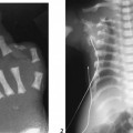
Radiologic Findings
Diagnosis
Discussion
Background
Etiology
Clinical Findings
Imaging Findings
RADIOGRAPHY
Related posts:


Stay updated, free articles. Join our Telegram channel

Full access? Get Clinical Tree


