 Clinical Presentation
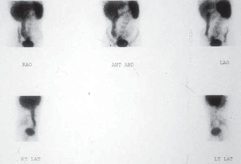
A 32-year-old woman with abdominal pain, fever, and diarrhea.

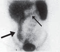
Multiple static images from a Tc99m-HMPAO (Ceretec) WBC study were obtained 30 minutes after tracer injection. Normal activity is seen within spleen, liver, bone marrow, and bladder. Abnormal WBC accumulation is seen to involve a long segment of the right and transverse colon (arrows).
 Differential Diagnosis
The liver, spleen, and bone marrow normally accumulate Tc99m-HMPAO–labeled leukocytes. Normal excretion of technetium complexes are also seen within the bladder and bowel on later imaging (unlike WBCs labeled with indium 111-oxine, which should not normally show bladder/bowel excretion).
• Inflammatory bowel disease (IBD): Tc99m-HMPAO–labeled WBC activity is seen in a pattern corresponding to large bowel that is abnormal (on early imaging) and compatible with colonic inflammation or infection.
• Pseudomembranous colitis: Bowel infection cannot be reliably differentiated from inflammation on WBC scan. A history of prolonged antibiotic use would favor this diagnosis.
• Gastrointestinal bleeding: This can look similar to IBD if active. Tracer will move over time if sequential images are obtained.
 Essential Facts


Stay updated, free articles. Join our Telegram channel

Full access? Get Clinical Tree


