Case 9

 Clinical Presentation
A 53-year-old woman with difficulty urinating.
 Imaging Findings

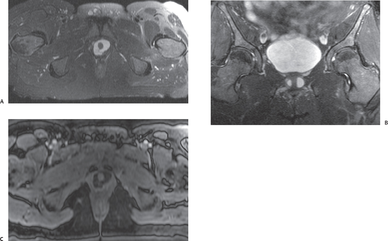
(A) Axial T2-weighted magnetic resonance imaging (MRI) of the lower pelvis shows a cystic structure (arrowhead) with contents of fluid signal intensity surrounding the proximal urethra (arrow). No filling defect is seen within this abnormality. (B) Coronal T2-weighted MRI of the pelvis confirms that the cystic abnormality (arrowheads) is located at the level of the urethra inferior to the urinary bladder (asterisk). The urinary bladder is normal. (C) Axial postcontrast fat-saturated T1-weighted MRI at the same level as Figure A shows no enhancement of the walls of the cystic structure (arrowhead). Note that the contents are of low signal intensity, consistent with simple fluid (asterisk).
 Differential Diagnosis
• Urethral diverticulum: This appears as a fluid-filled structure in the vicinity of or surrounding the urethra. The contents of the diverticulum show low signal on T1-weighted images and fluid signal intensity on T2-weighted images. A mildly enhancing wall may be seen. However, there is no filling defect or mass.
• Vaginal cysts:
Stay updated, free articles. Join our Telegram channel

Full access? Get Clinical Tree


