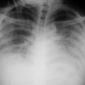
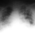
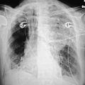
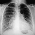
This is due to an area of pulmonary infarction secondary to the embolis. Remember, many cases of pulmonary emboli do not produce associated infarction. Effusions occur in about 50% of patients who have pulmonary infarcts, and they are generally associated with the patient’s developing chest pain. When an infarct has developed, this area does not tend to change rapidly, and one occasionally can distinguish among other causes of peripheral infiltrates due to edema or hemorrhage that do change fairly rapidly.
Fat embolism occurs when a large bone fractures and is also occasionally seen with pancreatitis, severe burns, or acute fatty liver. Neutral triglycerides are transported into the lungs where lipase hydrolyses into chemically active fatty acids that cause congestion, edema, intravascular coagulation, and, ultimately, alveolar hemorrhage. Chest x-rays are often normal in patients with small fat emboli, but with more extensive emboli a peripheral opacity can be seen. This occurs typically 1–3 days after trauma. There are other etiologies that cause pulmonary emboli, such as septic emboli from either an infected tricuspid valve or from intravenous drug injection. Also, occasionally, one can see amniotic fluid emboli from the uterus. Septic emboli most commonly are secondary to IV drug abusers or, occasionally, in immunodepressed patients. Patients with infected endocarditis on the tricuspid valve often develop small, multiple emboli throughout both lung fields. Staphylococcus aureus and streptococci are the organisms most commonly involved.
Stay updated, free articles. Join our Telegram channel

Full access? Get Clinical Tree


