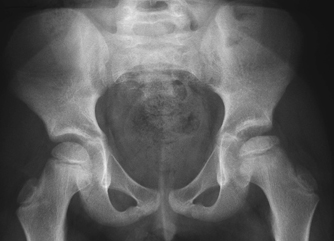
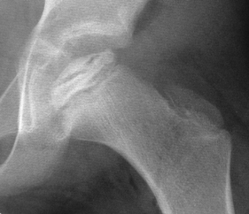
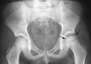
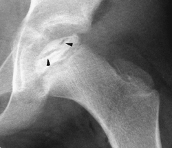
CASE 91 A 6-year-old male presents with a painless limp. Figure 91A Figure 91B A frontal view of the pelvis demonstrates a flattened inhomogeneous left femoral epiphyseal ossification center (Fig. 91A). A follow-up coned view of the left hip depicts an inhomogeneous flattened epiphyseal ossification center with areas of sclerosis and a large subchondral fissure (Fig. 91B) There is apparent widening of the medial joint space compartment. Legg-Calvé-Perthes disease (LCP) LCP is an osteochondrosis affecting the femoral head. The process is generally seen in Caucasian children 3 to 12 years of age, with a peak incidence between 4 and 8 years. Males are afflicted 5 times more frequently than females, with both hips involved in 10 to 15% of patients. Decreased birth weight, low socioeconomic class, attention-deficit disorder, delayed bone maturation, exposure to passive smoke, and abnormal birth presentation are all associated with an increased risk of LCP. A family history is seen in ~6% of cases. The origin remains uncertain; however, clotting abnormalities with resulting episodes of vascular thrombosis may play a role. Toxic synovitis, noted as a precursor for LCP in 1 to 3% of cases, however, is probably not a cause. Children classically present with a painless limp, although in the “synovitis phase” pain can be present, which may be referred to the knee. The pain is typically worse with activity and lessened with rest. Limitation of abduction and internal rotation of the hip, with resistance of “to and fro” rolling motion (+ log roll test) is found on physical examination. Atrophy of buttock, calf, and thigh musculature can be seen with long-standing disease. Leg-length discrepancy may be noted in more severe cases. Premature hip osteoarthritis may develop in the fourth or fifth decade, often necessitating hip arthroplasty. Osteochondritis dessicans develops in <5% of patients, often after a long symptom-free interval. Lateral extrusion of epiphysis can lead to “hinge” abduction, the result of failure of the femoral head to rotate medial with hip abduction due to blocking of the flattened femoral head on the lateral acetabular margin. Figure 91C AP radiograph of pelvis demonstrates a slightly flattened, sclerotic left femoral head epiphyseal center (arrows), which shows apparent mild lateral subluxation. Figure 91D Coned AP view of left hip depicts a subchondral fissure (arrowheads) spanning >50% of a slightly small, flattened sclerotic epiphyseal center. Four stages are typically described with all the osteochondroses:
Clinical Presentation
Radiologic Findings
Diagnosis
Differential Diagnosis/Consideration
Discussion
Background
Etiology
Clinical Findings
Complications
Pathology
Imaging Findings
RADIOGRAPHY
Related posts:


Stay updated, free articles. Join our Telegram channel

Full access? Get Clinical Tree


