 Clinical Presentation
A 74-year-old woman with newly diagnosed breast cancer. What is the most likely etiology for the appearance of the skull?

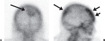
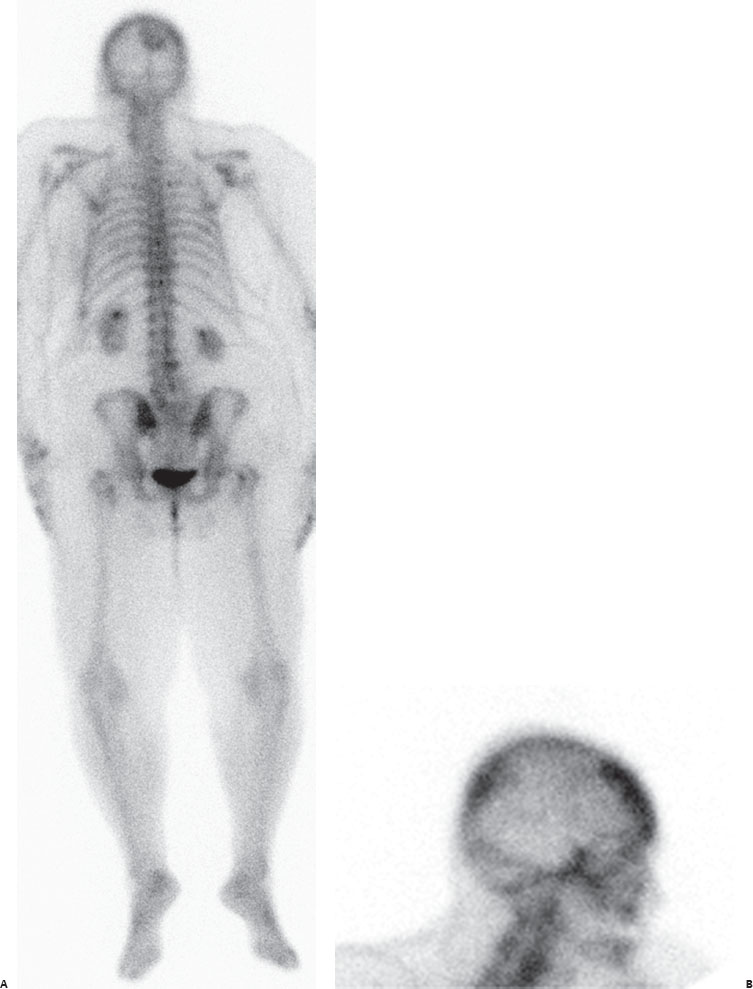
(A,B) Posterior image from a Tc99m-MDP whole-body bone scan reveals a large, round focus of increased tracer uptake in the right posterior parietal region (long arrow, A). This can be seen posteriorly on the right lateral spot view as well (short arrow, B). Tracer uptake in the frontal skull is typical for benign hyperostosis (double arrows, B). Degenerative uptake is seen in the right shoulder, lower lumbar spine, and hips.
 Differential Diagnosis
Tracer uptake in the region of the skull is difficult to evaluate on planar imaging because such uptake cannot always be differentiated from soft-tissue uptake in the adjacent brain.
• Intracranial soft-tissue uptake: This can be difficult to differentiate from bone uptake on planar images without SPECT. However, solitary skull region uptake is not uncommonly due to intracranial pathology (benign or malignant), which should be considered before the lesion is called a skull metastasis.


Stay updated, free articles. Join our Telegram channel

Full access? Get Clinical Tree


