 Clinical Presentation
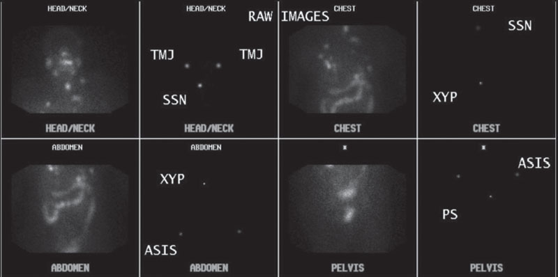
A 38-year-old woman with a history of malignancy presents for restaging.

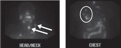
Anterior spot images from a whole-body (WB) scan with and without markers demonstrate multifocal uptake in the thyroid bed, lateral neck (arrows), and lung (circle). Physiologic activity is seen in the salivary glands, stomach, colon, and bladder.
 Differential Diagnosis
• Iodine 131 WB scan demonstrating metastatic thyroid cancer: Lack of normal bone uptake with physiologic salivary, gastric, intestinal, and bladder uptake makes radioiodine scan of metastatic thyroid cancer most likely.
• MIBG scan showing metastatic carcinoid: This has a somewhat similar biodistribution to iodine but typically also has normal myocardial uptake, and the tumor is more often below the diaphragm.
• Gallium scan showing metastatic melanoma:
Related posts:


Stay updated, free articles. Join our Telegram channel

Full access? Get Clinical Tree


