 Clinical Presentation
A 65-year-old man with left lung cancer presents for evaluation before a left pneumonectomy. His preoperative spirometry revealed a forced expiratory volume in 1 second of 1600 mL.

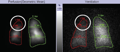
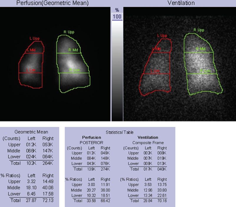
Posterior ventilation and perfusion images from a quantitative lung scan with 4 mCi of Tc99m-MAA and 10 mCi of Xenon 133. A matched ventilation–perfusion (VQ) defect is seen in the left upper lung corresponding to the known cancer on both ventilation and perfusion images (circles). A milder relative decrease is seen throughout the remainder of the left lung with a differential lung ventilation of 70% right and 30% left. Differential lung perfusion is 28% left and 72% right based on geometric mean analysis of anterior and posterior views (anterior images not shown).
 Differential Diagnosis
• Residual forced expiratory volume in 1 second (FEV1) of 1154 mL: After a left pneumonectomy, the residual FEV1 in this patient is predicted to be 1154 mL (1600 mL × 0.7213). This is calculated from the perfusion images with use of the geometric mean, which is the standard method recommended by the American College of Chest Physicians (ACCP). A postoperative FEV1 > 800 mL is considered adequate.
• Residual FEV1


Stay updated, free articles. Join our Telegram channel

Full access? Get Clinical Tree


