 Clinical Presentation
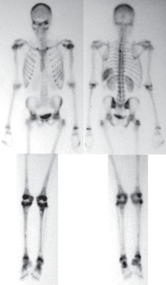
A 14-year-old boy with lower extremity pain.

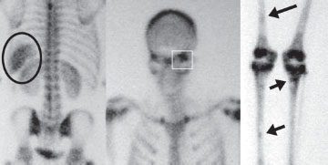
Three-hour–delayed anterior and posterior whole-body bone scan images demonstrate abnormal soft-tissue uptake in the left upper quadrant (circle). Also, increased activity is seen around the metaphyses of the knees and ankles and the sphenoid (square) that is due to marrow expansion. Other, subtler lesions of increased and decreased activity are seen in the extremities (arrows).
 Differential Diagnosis
• Sickle cell disease (SCD): Left upper quadrant soft-tissue uptake is consistent with autoinfarction of the spleen. Multifocal bony abnormalities (increased and decreased) are consistent with bony infarcts of various ages. The marrow expansion is also characteristic.
• Metastatic disease: This is unlikely as the majority of bone lesions are in the distal skeleton (beyond red marrow). Diffuse splenic activity is also very unlikely.
• Benign polyostotic bony process (e.g., polyostotic fibrous dysplasia or multiple enchondromas): Although either one of these benign bony processes could have similar bony appearances, they also do not have an effect on the spleen.
Related posts:


Stay updated, free articles. Join our Telegram channel

Full access? Get Clinical Tree


