 Clinical Presentation
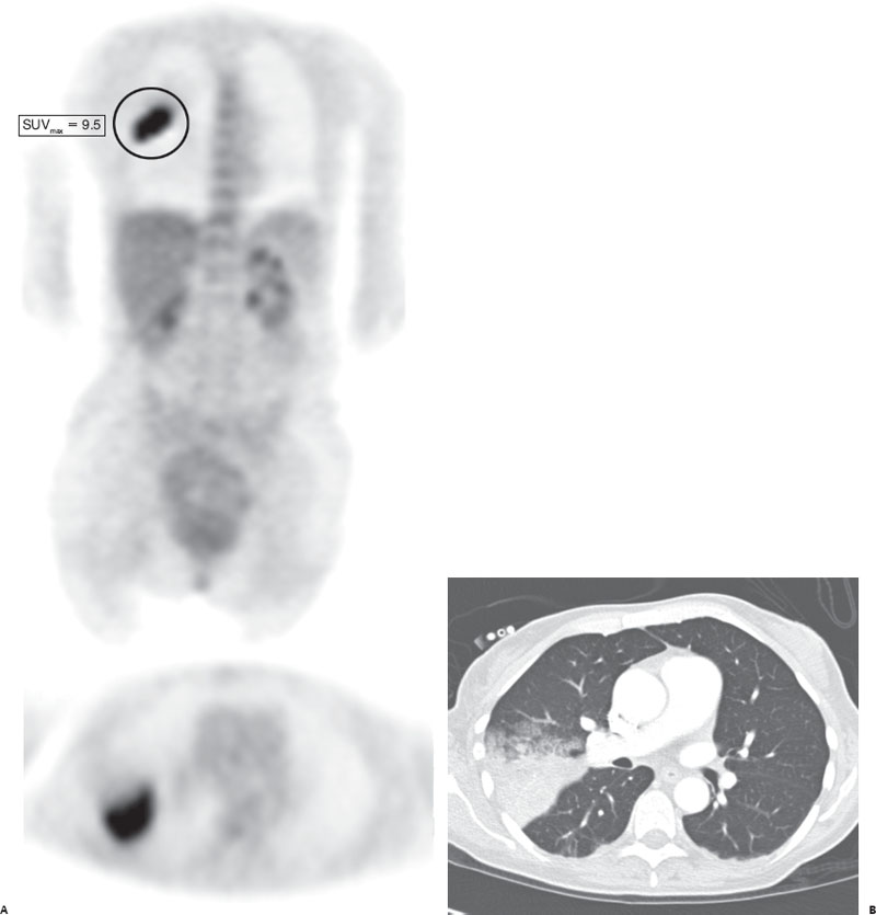
A patient presents for colon cancer restaging. The patient reports recent cough and fever.

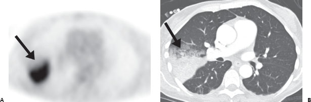
(A) Axial and coronal FDG-PET images demonstrate a markedly hypermetabolic lesion in the right mid lung (arrow). (B) Corresponding axial CT image demonstrates a wedge-shaped consolidative lesion that is sharply marginated posteriorly with adjacent ground-glass anteriorly (arrow).
 Differential Diagnosis
• Acute pneumonia: Segmental consolidation on CT is suggestive of infection, particularly given the history. Active infection will have increased FDG accumulation on PET.
• Synchronous lung carcinoma: This is a possible etiology. Follow-up chest radiography or CT should be performed after antibiotic treatment to demonstrate lesion resolution.
• Colon cancer metastatic to lung: This will also be hypermetabolic on FDG-PET but is typically more nodular on CT.


Stay updated, free articles. Join our Telegram channel

Full access? Get Clinical Tree


